All published articles of this journal are available on ScienceDirect.

Evaluation of Invasive Front and Depth of Invasion in Oral and Lip Squamous Cell Carcinoma and Clinicopathological Parameters

Authors Info & Affiliations
Abstract
Introduction
Oral Squamous Cell Carcinoma (OSCC) and Lip Squamous Cell Carcinoma (Lip SCC) are common head and neck malignancies with significant prognostic variability. Histological parameters, specifically the Invasive Front (IF) and Depth of Invasion (DOI), are critical in predicting tumor behavior and guiding treatment. This study examines IF and DOI in OSCC and Lip SCC and their correlation with tumor grade.
Methods
In this retrospective descriptive-analytical study, 80 patients (40 OSCC, 40 Lip SCC) treated at Ghaem and Omid Hospitals (Mashhad) and Razi Hospital (Tehran) from October 2022 to June 2024 were included. Biopsy samples with complete histopathological data were analyzed for IF, DOI, invasion pattern, vascular and neural invasion, and lymphocyte response. IF and DOI were classified as low or high risk, and tumor grading followed CAP guidelines. Data were analyzed using SPSS, with significance set at p < 0.05.
Results
In Lip SCC, 62.5% of cases were low-risk for IF and DOI, compared to 60% in OSCC. Lip SCC samples included 67.5% grade I, 20% grade II, and 12.5% grade III tumors, while OSCC showed 57.5%, 20%, and 22.5%, respectively. No significant differences were observed between OSCC and Lip SCC regarding IF or DOI. Significant correlations were found between histological grade and both IF and DOI, as well as vascular invasion, whereas neural invasion showed no correlation.
Conclusion
IF and DOI are reliable, accessible histopathological markers for evaluating OSCC and Lip SCC. Despite similar risk distributions, differences in anatomical location, oral environment, and early detection likely influence prognosis and treatment response. Incorporating IF and DOI assessment into routine histopathology can enhance prognostic accuracy and guide clinical decision-making.
1. INTRODUCTION
Head and Neck Squamous Cell Carcinoma (HNSCC) is the 9th most common cancer worldwide, accounting for approximately 90% of all head and neck malignancies. HNSCC affects the squamous mucosa of the upper aerodigestive tract, including the lip and oral cavity, and constitutes ~90% of all head and neck cancers [1]. Lip and oral cavity cancers are the most frequent non-melanoma head and neck cancers, with around 350,000 new cases diagnosed annually [2]. Oral Squamous Cell Carcinoma (OSCC) can arise in various oral sites, most commonly the tongue, and major risk factors include tobacco use and alcohol consumption, particularly in individuals in the sixth and seventh decades of life [3, 4]. Based on national and international guidelines, the most effective current treatments include surgery, radiotherapy, chemotherapy, and immunotherapy. Despite major advances in diagnoses and therapy since the 1970s, overall survival rates have remained unsatisfactory [5, 6]. In contrast, Lip Squamous Cell Carcinoma (Lip SCC) is generally less aggressive, with Ultraviolet (UV) light exposure, fair skin, older age, male sex, and the lower lateral vermillion border being recognized risk factors [7-9].
The TNM method, established by the American Joint Committee on Cancer (AJCC), evaluates tumor size (T), lymph node involvement (N), and distant metastases (M), which are among the most important characteristics used for therapy and prognostic management [10]. Clinical grading of the illness based on TNM classification and tumor location is frequently used as the main criterion for determining prognosis and therapy options [11]. However, even if the location and stage are the same, there are wide variations in treatment response and prognosis for oral squamous cell carcinoma; some patients live for a long time while others quickly metastasize [11, 12]. Furthermore, according to Almangush et al. [13], the BD histologic grading approach was used, which solely assesses tumor budding and depth of invasion in the invasive front region. There is also a strong correlation between these two factors and prognosis [11, 14, 15]. Tumor budding (B) denotes the presence of a solitary cancer cell or a small cluster of fewer than five cancer cells observed at the invasive front. Depth of invasion (D) was quantified from the tumor's surface to its farthest point of invasion, categorized as low (<4 mm) or high (≥4 mm). The histologic grading system entails the following criteria: a score of 0 corresponds to a tumor with <4 mm depth of invasion and fewer than five buds at the invasive front; a score of 1 aligns with a tumor that has ≥4 mm depth of invasion and fewer than five buds at the invasive front, or a superficial tumor with <4 mm depth of invasion but exhibiting high tumor budding activity at the invasive front (≥5 buds); a score of 2 designates a tumor with ≥4 mm depth of invasion and high tumor budding activity at the invasive front (≥5 buds). Tumors with a score of 0 are categorized as low risk, those with a score of 1 as intermediate risk, and those with a score of 2 as high risk [15].
Strieder et al. [15] compared some histologic grading methods for squamous cell carcinoma of the lip. They discovered that combining TNM staging with the BD model of histologic grading can aid in treatment planning, preventing mutilations and the resulting loss of patient quality of life. According to them, the BD model was the most objective, straightforward, and useful technique for determining the prognosis of lower lip SCC.
It is critical to select the right treatment technique since any flaw may harm the patient in the long run, whether in the form of recurrence, residual lesion, or irreversible morbidity. Many recent studies have demonstrated that if the initial therapy fails, the disease-related mortality rate skyrockets, making survival a distant possibility [15]. This retrospective study aims to compare Invasive Front (IF), Depth of Invasion (DOI), and other histomorphologic parameters between intraoral SCC and lip SCC.
2. MATERIAL AND METHOD
2.1. Patients and Tumors
This retrospective descriptive analytical study was conducted on 80 biopsy samples, including 40 tumor tissues of intraoral squamous cell carcinoma and 40 tumor tissues of lip squamous cells, resulting from complete surgery of lesions and with healthy surgical margins. Samples were collected from the Pathology Departments of Razi Skin Hospital, Tehran, and Omid and Ghaem Hospitals, Mashhad, Iran. Biopsy samples collected between October 2022 and June 2024 were included in this study. Demographic information of patients, including age, sex, and lesion history, was recorded. This retrospective study was conducted in accordance with the Declaration of Helsinki. Ethical approval was obtained from the Institutional Review Board of Mashhad University of Medical Sciences (Approval number: IR.MUMS.DENTISTRY.REC.1400.330). Patient data were anonymized. Available OSCC biopsy samples were included from samples with adequacy to examine IF and DOI. Inclusion criteria comprised adequate tissue for IF and DOI evaluation, confirmed histopathological diagnosis of OSCC or Lip SCC, complete pathology files, no prior anti-tumor treatment, and healthy surgical margins. Exclusion criteria included oxygen biopsy, incomplete files, low-quality slides, disagreement between pathologists on diagnosis, and unavailable slides/blocks.
2.2. Histopathology and Histomorphology Factors
In the studied samples, the variable depth of invasion and the invasive edge were examined and measured by a pathologist with an optical microscope without knowing the clinical information of the patients. To check the invasive edge, the samples were first examined at 40 times magnification, and then the areas with the most budding were selected. Cells were counted with 400 times magnification, and the highest value in each sample was recorded as the score. Then the samples with the number of larger cells equal to 5 are classified as high risk group, and less than 5 cells are classified as low risk group [16]. All paraffin blocks and slides were standardized following a uniform sectioning protocol. Serial sections of 4 µm thickness were prepared, and representative tumor areas were selected for IF and DOI analysis. Two experienced pathologists evaluated all samples independently, blinded to the patients’ clinical data and tumor site. In cases of disagreement, consensus was reached through joint review.
The Depth of Invasion (DOI) was measured from the basement membrane of the adjacent intact mucosa to the deepest point of tumor infiltration. Based on a 4-mm cutoff, patients were categorized into high-risk (DOI > 4 mm) and low-risk (DOI < 4 mm) groups [16]. Cellular pleomorphism was assessed in tumor sections by estimating the percentage of pleomorphic cells per High-Power Field (HPF). The scoring system applied was as follows: (1) 0–5%, (2) 6–25%, (3) 26–50%, and (4) > 50%. The host lymphocytic response at the deepest invasive front was also evaluated and classified into four grades, such as marked (> 50%), moderate (25–50%), mild (5–25%), and absent (0–5%) [17]. The pattern of tumor invasion into the connective tissue was recorded and categorized into three types, including pushing, group, and strand patterns [18]. Perineural invasion and vascular invasion were assessed by determining the presence or absence of tumor cells around nerves, and around or within blood vessels.
Subsequently, DOI, invasive front pattern, and lymphocytic response were compared between intraoral SCC and lip SCC. Pairwise comparisons between groups were performed using the chi-square test and the t-test, where appropriate.
2.3. Statistical Analysis
The sample size for each group (OSCC and Lip SCC) was calculated using PASS software, with 95% confidence level and 90% statistical power based on expected differences in histological grades. The minimum required sample size was 33 per group, which was increased to 40 per group to enhance reliability. Data were analyzed using SPSS software version 24 (IBM Corp., Armonk, NY, USA). Continuous variables were presented as mean ± Standard Deviation (SD), and categorical variables were expressed as frequencies and percentages. The chi-square test was applied to compare categorical variables such as Invasive Front (IF) risk, Depth of Invasion (DOI) risk, and histological grade between groups. The independent t-test was used to compare continuous variables, such as age. A p-value <0.05 was considered statistically significant. All analyses were performed by a blinded statistician.
3. RESULTS
A total of 80 patients were included, comprising 40 cases of Lip SCC and 40 cases of OSCC. The demographic characteristics are summarized in Table 1. There was no statistically significant difference between the two groups regarding age or gender distribution (p > 0.05).
| Characteristic | Lip SCC (n=40) | OSCC (n=40) | Chi-square | P-value |
|---|---|---|---|---|
| Gender | 2.581 | 0.108 | ||
| Female | 12 (30%) | 19 (47.5%) | ||
| Male | 28 (70%) | 21 (52.5%) | ||
| Age | 2.619 | 0.270 | ||
| <50 years | 6 (15%) | 7 (17.5%) | ||
| 50–70 years | 23 (57.5%) | 16 (40%) | ||
| >70 years | 11 (27.5%) | 17 (42.5%) |
3.1. Invasive Front and Depth of Invasion
Analysis of the invasive front showed that 62.5% of Lip SCC cases and 60% of OSCC cases were classified as low-risk, while 37.5% and 40% were high-risk, respectively. Similar proportions were observed for depth of invasion, with no statistically significant differences between the two groups (p = 0.818; Table 2).
| Parameter | Risk Category | Lip SCC (n=40) | OSCC (n=40) | Chi-square | P-value |
|---|---|---|---|---|---|
|
Invasive Front (IF) |
Low risk | 25 (62.5%) | 24 (60%) | 0.53 | 0.818 |
| High risk | 15 (37.5%) | 16 (40%) | |||
|
Depth of Invasion (DOI) |
Low risk | 25 (62.5%) | 24 (60%) | 0.53 | 0.818 |
| High risk | 15 (37.5%) | 16 (40%) |
3.2. Pattern of Invasion
In Lip SCC, the push pattern was most common (50%), followed by group (35%) and strand (15%). In OSCC, 52.5% showed a push pattern, 27.5% a group pattern, and 20% a strand pattern. No significant difference was found between the two groups (p = 0.715).
3.3. Vascular and Perineural Invasion
Vascular invasion was observed in 37.5% of Lip SCC cases and 47.5% of OSCC cases. Perineural invasion occurred in 22.5% and 10% of cases, respectively. Neither variable showed a statistically significant difference between groups (p = 0.366 and p = 0.13, respectively).
3.4. Histological Grade and Its Associations
In Lip SCC, 67.5% of cases were grade 1, 20% grade 2, and 12.5% grade 3. In OSCC, 57.5% were grade 1, 20% grade 2, and 22.5% grade 3.
Histological grade was significantly associated with vascular invasion (p < 0.01), with grade 3 observed more frequently in samples showing vascular invasion. No significant association was found between perineural invasion and tumor grade (p = 0.113).
A significant association was observed between invasive front risk category and histological grade, with all grade 3 tumors classified as high-risk (p < 0.01). Depth of invasion was also significantly correlated with histological grade (p < 0.05), with high-risk DOI more frequently observed in grade 3 tumors (Fig. 1).
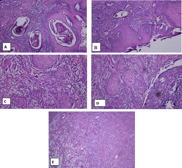
Histopathological features of Lip Squamous Cell Carcinoma (Lip SCC). (A) Grade 1 Lip SCC with well-differentiated tumor islands. (B) Invasion pattern of the tumor front (pushing pattern). (C) Moderate Lymphocytic Host Response (LHR) at the invasive front. (D) Moderate cellular polymorphism. (E) Invasive Front (IF) and Depth of Invasion (DOI) assessment: low-risk IF and high-risk DOI. All images were evaluated according to CAP guidelines. Scale bars = X μm.
4. DISCUSSION
OSCC and Lip SCC are among the malignant lesions of the head and neck region, and these two lesions, despite morphological similarities, differ in terms of prognosis and survival. Considering the unpredictable behavior of OSCC in the digestive and respiratory systems, the prognosis of lip SCC is much better than that of OSCC. Cancer prognosis is described by the invasive front of cancerous cells into the stroma and vessels, their depth of invasion, histopathological features, colonization, and proliferation in the lymph nodes. Recent studies show that the number of cells at the invasive front can be a prognostic factor in early-stage patients, with more than 5 cells at the edge of invasion indicating a poorer prognosis. According to studies, the depth of invasion of 4 mm or more is strongly related to the possibility of cervical metastasis, and its increase is strong along with the increase in mortality in patients with oral SCC [19]. Our study aimed to evaluate and compare invasive front, depth of invasion, and CAP grading indices in oral SCC and Lip SCC.
This study found that the relationship between the invasive front and histological differentiation is significant. This measure was also significant for the depth of invasion. It was found that there is a significant relationship between vascular invasion and histological differentiation, but this criterion was not significant for neural invasion. Also, according to the significant relationship between the lymphocyte response and the degree of the disease, it can be concluded that the lower the lymphocyte response, the higher the degree of the disease.
In addition to histopathological parameters, the oral mucosal microenvironment plays a crucial role in modulating tumor invasion and progression. Chronic irritation, mechanical trauma, and the presence of orthodontic appliances or other prosthetic devices can induce local inflammatory responses, leading to alterations in tissue architecture and stromal composition. These changes may promote epithelial-mesenchymal transition and facilitate cancer cell invasion, thereby influencing both the Invasive Front (IF) and Depth of Invasion (DOI) in OSCC and lip SCC. The inflammatory microenvironment, characterized by increased lymphocyte infiltration, cytokine release, and vascular changes, can modify the tumor-stroma interactions and affect histological patterns observed at the invasive edge. Therefore, considering the impact of local mucosal factors is essential for interpreting IF and DOI findings, as these microenvironmental influences may contribute to variability in histological differentiation, tumor aggressiveness, and ultimately patient prognosis.
In the 2021 study by Mohtsham et al., 87 OSCC cases were evaluated and classified into low-risk and high-risk groups based on the pattern of the invasive front. Overall, 50.6% of the cases were categorized as low-risk and 49.4% as high-risk. Notably, 75% of patients in the low-risk group presented with grade 1 tumors [20]. In the current study, in the examination of the edge of invasion, 62.5% of lip SCC samples were in the low-risk group, and 37.5% were in the high-risk group, and in the OSCC samples, 60% were low risk, and 40% were high risk, which is consistent with the results of the above study.
In a study by Wang et al. in 2011, tumor buddings were counted. Those who had more than or equal to 5 buds were placed in the high intensity group. Their results show that among 230 patients, 111 samples were in the high intensity group. Their study showed that the number of buds is related to the size of the tumor and the degree of differentiation [21]. In our study, IF status was also significantly related to histological differentiation; So that the low-risk group is better differentiated, included grade 1 and 2.
In the study by Yuen et al., which included 72 patients with tongue carcinoma, the invasive edge grading method introduced by Byrne and tumor thickness were used. The samples were divided into three groups by thickness: less than or equal to 3 millimeters, 3 to 9 millimeters, and greater than 9 millimeters. They found that 59.7% of patients were in the middle group in terms of thickness [22]. In our study, DOI was evaluated, which, according to the CAP protocol, has greater prognostic value than tumor thickness.
Almangush et al.'s study in 2020 [19] aimed to investigate tumor depth and the invasive front in 233 early-stage OSCC patients. They evaluated tumor depth from the tumor edge to the deepest point of invasion and divided it into two groups: greater than or equal to 4 mm or less than 4 mm. Among the patients, 153 people (65.6%) were placed in the high group. In the two groups of our study, 40% and 37.5% of the cases were in the high group. This difference may be due to sample size, racial and geographical differences, and oral habits.
P. Santos et al. investigated grading systems in squamous cell carcinoma of the lip in 2014. In this study, which included 59 patients with lip SCC, the samples were also examined for invasion pattern. In terms of invasive front grading, patients were divided into low and high groups. Sixty-one percent of patients were in the high-grade group of invasive edge. In terms of invasion pattern, the patients were divided into five groups: (1) pushing, (2) large separated islands, (3) small tumor islands, (4) tumor satellite, and (5) finger. Overall, 52.5% of patients were placed in groups 1, 2, and 3 in terms of invasion pattern [23]. Despite differences in the classification of the invasion pattern, 65% of the samples we studied in the PUSH and STRAND groups were almost similar to the above study.
It can be concluded that the significant relationship between histological differentiation and IF status indicates that this is the best way to evaluate the invasion rate in lip SCC and OSCC. Parameters related to IF and DOI are cheaper and more accessible tools than biological markers for evaluation the new grading of SCC. The lack of statistically significant difference between the new grading indices in lip SCC and OSCC indicates that the location of lip SCC and early detection of the lesion compared to oral SCC, the difference in the biological conditions of the oral environment compared to vermilion, oral habits, anatomical proximity such as the distance from glands Lymphatic and epigenetic factors effective in saliva, can be one of the main factors affecting prognosis and response to lesion treatment in these two adjacent places.
The grading of the studied samples was done based on the criteria of the CAP protocol, and for the first time, lip SCC and OSCC were compared from the perspective of new grading methods. Our most important limitation in conducting this study was that some low-quality samples were excluded, and it was difficult to obtain replacement samples due to the limited number available. Incompleteness of patients' files in medical-educational and research centers caused the withdrawal of a significant number of samples from the study.
5. LIMITATION AND FUTURE DIRECTIONS
This retrospective, cross-sectional comparative study provides valuable insights into the prognostic significance of Invasive Front (IF) and Depth of Invasion (DOI) in Oral and Lip Squamous Cell Carcinoma (OSCC and Lip SCC). However, several limitations should be considered. The strict inclusion criteria and limited availability of high-quality biopsy samples resulted in a total of 80 patients, which may restrict the generalizability of the findings. Incomplete medical records led to the exclusion of some patients, potentially affecting the accuracy of correlations between histological parameters and clinical outcomes. Moreover, variability in tissue handling and slide quality could introduce minor biases in pathological assessment.
Future research should focus on prospective, multicenter studies with larger and more diverse patient populations to reduce selection bias and improve generalizability. Standardized protocols for sample collection, slide preparation, histopathological evaluation, and data management are recommended to minimize variability and ensure reproducibility of IF and DOI measurements. These studies could further clarify the clinical implications of IF and DOI in surgical decision-making and postoperative follow-up strategies for OSCC and Lip SCC.
CONCLUSION
Considering the significant relationship between histological differentiation and both IF and DOI, these parameters are valuable for evaluating the invasion pattern and prognosis of oral and lip SCC. Moreover, IF and DOI can directly inform clinical decision-making by guiding the determination of surgical margins, risk stratification for recurrence, and the design of postoperative follow-up protocols. These findings highlight the practical relevance of histopathological assessment in patient management, providing accessible and cost-effective tools compared to molecular markers. Further studies integrating IF and DOI with patient outcomes could refine surgical and therapeutic strategies in OSCC and lip SCC.
AUTHORS’ CONTRIBUTIONS
The authors confirm their contribution to the paper as follows: F.M., N.M.: Study conception and design; F.M.: Funding acquisition; F.M., S.A.: Project administration; S.A., N.E.: Investigation / Data collection; K.D., S.A.: Data curation; M.Sh.: Formal analysis and statistical analysis; K.D., N.E.: Manuscript drafting; F.M., S.A., N.M., K.D., N.E., M.Sh.: Review & editing; F.M., N.M.: Supervision. All authors have read and approved the final manuscript and are accountable for all aspects of the work. The authors confirm that the contents of this manuscript have not been published nor submitted elsewhere.
LIST OF ABBREVIATIONS
| OSCC | = Oral squamous cell carcinoma |
| Lip SCC | = Lip squamous cell carcinoma |
| IF | = Invasive front |
| DOI | = Depth of invasion |
ETHICS APPROVAL AND CONSENT TO PARTICIPATE
Ethical approval was obtained from the Institutional Review Board of Mashhad University of Medical Sciences, Iran (Approval number: IR.MUMS.DENTISTRY.REC.1400. 330).
HUMAN AND ANIMAL RIGHTS
All procedures performed in studies involving human participants were in accordance with the ethical standards of institutional and/or research committee and with the 1975 Declaration of Helsinki, as revised in 2013.
AVAILABILITY OF DATA AND MATERIALS
The data supporting this study's findings will be made available from the corresponding author [F.M.] upon reasonable request.
FUNDING
This study was funded in full by Mashhad University of Medical Sciences, Faculty of Dentistry, Mashhad, Iran, with the grant number 991611.
ACKNOWLEDGEMENTS
All authors thank the Department of the Research Council of Mashhad University of Medical Sciences, Faculty of Dentistry, Mashhad, Iran.