All published articles of this journal are available on ScienceDirect.

Gingival Overgrowth in an E-Cigarette User: A Case Report of a Possible Association

Abstract
Background
The use of electronic cigarettes (e-cigarettes) has significantly increased among young adults, yet the effects on oral health remain inadequately understood. Emerging evidence suggests a potential link between vaping and periodontal inflammation, as well as alterations in oral mucosa, but there is limited clinical case documentation.
Case Presentation
A 26-year-old male, a regular e-cigarette user with poor oral hygiene, presented with a localized, erythematous gingival mass located between the maxillary right lateral incisor and canine (#12–#13). The lesion had a smooth surface and bled easily upon manipulation. Initial management included nonsurgical periodontal therapy, reinforcement of oral hygiene practices, and smoking cessation counseling. Due to the persistence of the lesion, a surgical excision was performed that extended beyond the visible margins. Histopathological examination revealed a benign reactive lesion characterized by acanthosis, fibrosis, and chronic inflammatory infiltrate, with no evidence of malignancy. Follow-up at six weeks demonstrated satisfactory healing with no recurrence.
Conclusion
This case underscores a potential association between e-cigarette use and reactive gingival lesions. Persistent gingival enlargements in patients who vape should not be overlooked and require thorough evaluation, including a biopsy when necessary. A comprehensive patient history, elimination of local irritants, and long-term follow-up are crucial. Further studies are needed to clarify the causal relationship between vaping and oral soft tissue pathology.
1. INTRODUCTION
Young adults' use of e-cigarettes has grown significantly, and most of these people believe that e-cigarettes are less harmful compared to the usual method of smoking [1]. Public health institutions continue to inform the public that the substances in e-cigarettes are not safe and should not be considered safe, as they may have effects on various parts of the body, including the mouth [2].
Nicotine, heated aerosol, solvents (propylene glycol/vegetable glycerin), and flavorings are directly delivered into the oral cavity [3]. Symptoms of periodontitis may be initiated or exacerbated by alterations in the oral environment, including changes in salivary composition, tissue irritation, and inflammatory responses. Although evidence regarding the oral effects of electronic cigarettes is still emerging, current literature suggests an association between e-cigarette use and adverse periodontal outcomes, including gingival inflammation and the progression of periodontitis [4].
Calculus, plaque, and other locality-related conditions have major effects on periodontal diseases and reactive gingival enlargements. These are common causes of reactive soft tissue masses that may manifest as other diseases, and they are also major causes of erythema, edema, and bleeding on probing. Therefore, it is important to differentiate between plaque-induced lesions and other conditions commonly encountered in dentistry [5].
In cases where a gingival lesion is persistent, it may be challenging to differentiate its nature through clinical observation alone. Some reactive lesions might seem relatively harmless, as is the case with inflammatory hyperplasia. However, if a lesion persists even after the offending irritant has been removed, or appropriate periodontal treatment has been administered, a biopsy may be necessary to rule out dysplasia or carcinoma [6].
Potential oral symptoms in e-cigarette users, such as increased gingival bleeding, periodontal inflammation, and altered oral microbial profiles, have been the subject of more and more published reports and reviews in the field of dentistry. Despite ongoing research, the evidence remains conflicting and developing. Many clinicians may not consistently consider vaping as a contributing risk factor, especially when traditional irritants like plaque are present [7, 8].
This report presents the clinical course, management approach (initial debridement and hygiene reinforcement followed by excision), and histopathologic outcome (benign reactive changes with acanthosis, fibrosis, and chronic inflammation) to substantiate clinical decision-making in similar cases. It emphasizes the need for a comprehensive history of exposure, including details about vaping, control of local irritants, and biopsy of persistent gingival masses for proper diagnosis and follow-up.
2. CASE REPORT
2.1. Case Presentation
A 26-year-old male visited the dental clinic on May 24, 2023, with a primary complaint of localized swelling in the gums in the maxillary anterior region. The patient reported a history of regular electronic cigarette (e-cigarette) use and acknowledged poor oral hygiene practices. His medical history was otherwise unremarkable.
During the clinical examination, it was observed that the lesion had gradually increased in size over an unspecified period and was occasionally associated with bleeding while brushing. The patient did not report any pain or systemic symptoms.
2.2. Clinical and Radiographic Examination
An intraoral examination revealed a localized, erythematous gingival mass situated on the buccal aspect between the maxillary right lateral incisor and canine (#12–#13). The lesion appeared smooth-surfaced and sessile, with bleeding upon gentle manipulation (Fig. 1).
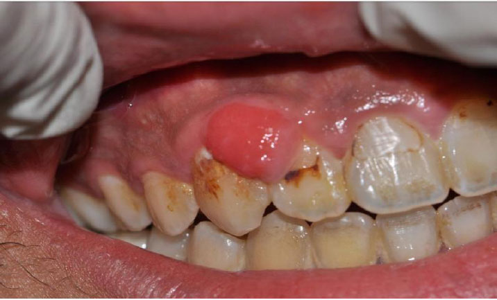
Pre-operative clinical view. Raised erythematous gingival mass between #12 and #13 with plaque deposits; lesion shows a smooth surface and clinical tendency to bleed.
A periodontal assessment indicated plaque accumulation and signs of poor oral hygiene. No significant tooth mobility was detected in the affected area.
Radiographic evaluation using a periapical radiograph revealed no underlying bone loss, root resorption, or associated radiolucent/radiopaque lesions around the affected teeth, indicating that the lesion was confined to the soft tissue.
2.3. Treatment Plan and Management
The initial management focused on eliminating local factors contributing to the condition through nonsurgical periodontal therapy (Fig. 2). This included scaling and root debridement, along with comprehensive oral hygiene instructions. The patient was also advised to stop using e-cigarettes and was provided with counseling to assist with cessation (Table 1).
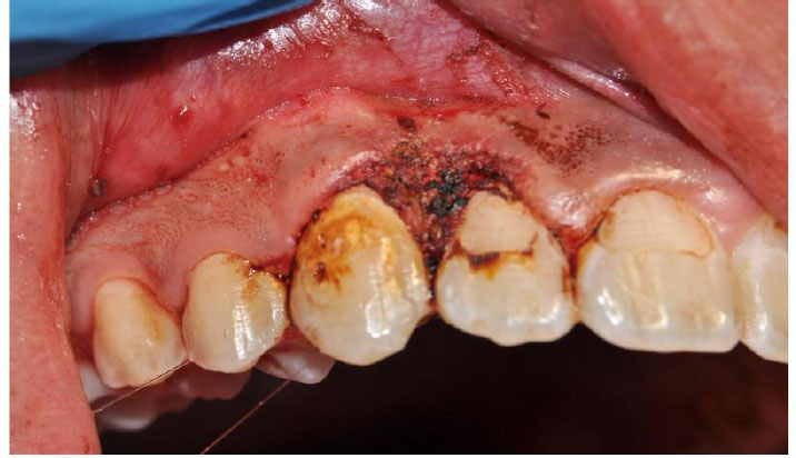
Day of surgery. Immediate post-excision surgical field after removal beyond visible margins.
| Time | Event |
|---|---|
| May 2023 | Presentation |
| Initial visit | Scaling + OHI |
| After persistence | Surgical excision |
| 6 weeks | Healing |
Despite initial treatment, the lesion persisted with no significant improvement. Consequently, the decision was made to proceed with surgical excision.
The lesion was removed with an adequate margin beyond the visibly affected areas to ensure complete excision. The excised tissue specimen was then sent for histopathological examination to rule out any dysplastic or malignant changes (Fig. 3).
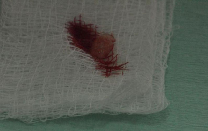
Biopsy specimen. Excised tissue shown on gauze and clinical site image taken around biopsy collection.
The histopathological examination of the removed gingival tissue revealed a 0.9 × 0.5 cm tan soft tissue fragment. Microscopic examination was consistent with a benign/reactive process, acanthosis, and underlying fibrosis with chronic inflammation. It did not indicate any malignant changes (Fig. 4).
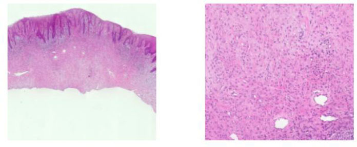
Histopathology. Representative sections consistent ith acanthosis and chronic inflammatory changes with underlying fibrosis.
Postoperative healing was smooth, and clinical follow-up at six weeks showed satisfactory healing, with no signs of recurrence or residual inflammation (Fig. 5).
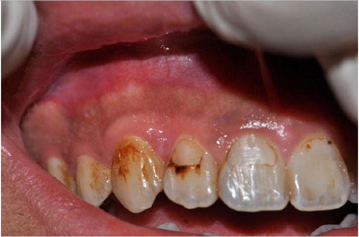
Follow-up after 6 weeks. Healed gingival tissue at the excision site with no visible inflammatory recurrence.
3. ETHICAL CONSIDERATION
The clinical records, radiographs, and histopathological data were obtained during the patient’s routine clinical care. Informed consent for the publication of the patient’s clinical information and images was obtained and signed by the patient.
4. DISCUSSION
There has been a significant increase in e-cigarette use among young adults, yet there is insufficient information regarding its oral manifestations, particularly regarding soft tissue lesions presenting with features of reactive and potentially malignant lesions. The current case presentation involves a 26-year-old regular e-cigarette smoker who presented with an erythematous mass over the right maxillary region between the lateral incisor and canine (#12-#13 regions), which bled easily on touch and was found to be benign with fibrosis, acanthosis, and chronic inflammation on histopathological evaluation and also failed to recur at 6-week follow-up post- excisional biopsy. Its clinical significance lies in the fact that it signifies that exposure to e-cigarettes is linked with probable reactive and inflammatory enlargement of the gingiva, giving rise to neoplasia-related features and hence is fit for biopsy.
The results of systematic reviews have shown that e-cigarette aerosols are detrimental to the oral mucosa, as evidenced by symptoms such as mucosal irritation, periodontal/gingival alterations, and other effects [9]. The adverse effects of e-cigarette exposure on oral components can be categorized into several categories, pointing out that gingival inflammation, along with other adverse effects on the mouth, has been prominent. Ralho et al. (2019) indicated that e-cigarette smoking increases vulnerabilities to alterations of the mouth and periodontal regions, noting adverse effects on mucosa such as mucosal lesions, nicotinic stomatitis, hairy tongue, angular cheilitis, and prominent inflammation [10]. The study by Holliday et al., 2021, underscored that while there have been very few clinical cases to prove adverse effects of e-cigarette smoking on the mouth, adverse effects such as dryness of the mouth, irritation, and gingivitis have been prominent, according to epidemiological research [11]. The evidence demonstrates a consistent correlation with clinical manifestations, such as cutaneous flushing and bleeding tendencies. Additionally, there is histopathological evidence of chronic inflammation and acanthosis (thickening of the epithelial layers due to chronic irritation). This suggests that the observed changes may be a reactive response rather than indicative of a neoplastic process.
E-cigarette aerosols contain substances such as nicotine, humectants (propylene glycol and vegetable glycerin), and flavoring agents. These components can directly interact with the genetically distinct gingival epithelial and connective tissues in the mouth. Based on previously reviewed pathophysiological explanations and mechanisms, these exposures are believed to contribute to oxidative stress and inflammation. They may also lead to dysbiotic changes in the interactions between the host and the microbiome, as well as impair the repair processes of these tissues.
Javed et al. (2017) presented mechanistic explanations and discussions outlining how certain components found in e-cigarette aerosols may contribute to the formation of reactive oxygen species and carbonyl stress responses. These processes can lead to inflammation and disrupt repair mechanisms within periodontal tissues [12]. Wilson et al. (2022) provided additional explanations and summaries of several cellular phenomena associated with exposure to e-cigarette aerosols in head and neck/oral cells. These phenomena include cytotoxicity, oxidative stress, decreased cell viability, delayed fibroblast migration, and genotoxicity. Such effects may contribute to conditions like chronic mucositis and hyperplasia [13].
Recently, Ganapathy et al. (2024) discussed how components in aerosols can collectively elevate reactive oxygen levels, diminish antioxidant defenses, cause DNA damage, and trigger the release of pro-inflammatory cytokines. These mechanisms may help explain the histopathological triad observed in patient fibrosis, acanthosis, and chronic inflammation, indicating a reactive lesion possibly linked to aerosol irritation [14].
A key clinical message from this case is the need for a cautious diagnostic approach. Gingival masses that are erythematous, bleed easily, or appear to enlarge can overlap in appearance with reactive lesions (e.g., inflammatory hyperplasia) as well as premalignant or malignant entities. Because e-cigarette aerosols have been associated with cytotoxic/genotoxic properties and because long-term clinical risks remain uncertain, surgical excision and histopathologic assessment as performed here remain appropriate when clinical features are atypical or when malignancy cannot be excluded. Yang et al. (2020) [9] and Holliday et al. (2021) [11] both underscore the ongoing uncertainty and variability across products and users, reinforcing the need for clinicians not to assume benignity solely on the basis of patient age or vaping history.
Complete excision with extension beyond visible margins resulted in satisfactory healing and no recurrence at 6 weeks. While short-term outcomes are reassuring, the literature emphasizes that clinical studies on oral outcomes of vaping are often small and cross-sectional, and long-term lesion behavior is not well defined. With e-cigarette aerosols being reported as having potential cytotoxic/genotoxic activity and with clinical long-term risk being uncertain, surgical excision with histopathological analysis, as done in this case, can be justified whenever clinical presentation is suspicious or whenever malignancy is not ruled out. This has been well discussed by Yang et al., 2020 [9], and further confirmed by Holliday et al., 2021 [11], who state that clinical experience should not be misled by age and vaping experience alone.
Causality cannot be established from a single case; local factors (plaque accumulation, trauma, or undetected irritants) might also contribute to gingival reactive lesions. Poor oral hygiene and plaque-induced inflammation were also considered possible contributing factors; therefore, the patient received professional periodontal cleaning and oral hygiene instructions. The temporal association with regular vaping, the lesion's inflammatory clinical phenotype, and the concordant histopathology provided a clinically useful signal that was consistent with available evidence that vaping may be associated with oral mucosal and gingival inflammation. This report further supports the growing clinical awareness that cigarette use may be associated with peculiar oral soft tissue reactions in young adults and confirms the continuation of recommendations for biopsy of suspicious gingival enlargements and for targeted preventive counseling in users of e-cigarettes.
5. LIMITATIONS
This report is limited by its single-patient design, which precludes establishing a causal relationship between e-cigarette use and the gingival lesion. Local factors, particularly plaque accumulation and plaque-induced inflammation, as well as mechanical trauma or unidentified irritants, may have contributed to the lesion. Although professional periodontal cleaning and reinforcement of oral hygiene were provided and the lesion persisted despite these measures, the contribution of such confounding factors cannot be completely excluded. Further clinical studies involving larger patient populations are required to clarify whether e-cigarette exposure plays a specific role in the development of reactive gingival lesions.
CONCLUSION
This case report represents a possible association between e-cigarette use and oral lesions, specifically reactive gingival lesions. Since the lesion did not improve with nonsurgical periodontal treatments and enhanced oral hygiene, it was surgically removed and sent for histopathological testing. The results confirmed that it was a benign reaction, with no signs of malignancy found. During the follow-up examination conducted six weeks later, there was evidence of complete healing, indicating that no recurrence occurred within that time frame. This case emphasizes the importance of further investigating the potential link between vaping and benign gingival reactions. It also highlights the need for a thorough evaluation of patients who vape, including education on oral hygiene and strategies for quitting, as well as biopsy for any remaining gingival lesions after the source of irritation has been removed.
AUTHOR’S CONTRIBUTION
The author confirms sole responsibility for the following: H.A.: Study conception and design, data collection, analysis and interpretation of results, and manuscript preparation.
AVAILABILITY OF DATA AND MATERIALS
The data supporting the findings of the article will be available from the corresponding author [H. A] upon reasonable request.
FUNDING
This study is supported via funding from Prince Sattam bin Abdulaziz University project number (PSAU/2025/R/ 1447)
ACKNOWLEDGEMENTS
The authors would like to thank Image Dental Clinic for their valuable support in facilitating the clinical procedures and patient management involved in this case report.