All published articles of this journal are available on ScienceDirect.

From Biologic Width to Biologic Belt: A Three-dimensional Conceptual Framework for Supracrestal Tissue Attachment

Editorial
Background
The concept of biologic width has long been used to describe the supracrestal soft tissue dimensions surrounding natural teeth and to guide restorative and periodontal procedures. Traditionally, this concept has been interpreted as the vertical dimension of the junctional epithelium and supracrestal connective tissue attachment measured from the alveolar bone crest.
Conceptual Gap
Despite its clinical utility, biologic width has traditionally been interpreted primarily as a vertical linear measurement, which may not fully emphasize the circumferential three-dimensional organization and biological function of the supracrestal tissues.
Proposed Concept
This article proposes a novel conceptual framework, termed the biologic belt, that complements the established concepts of biologic width and supracrestal tissue attachment (STA) by emphasizing their continuous, circumferential, and three-dimensional protective organization. Beyond its geometric description, the term “belt” highlights the protective role of this tissue complex, a three-dimensional biological barrier safeguarding periodontal tissues against mechanical, microbial, and restorative challenges.
Clinical Relevance
Reconceptualizing the supracrestal tissues as a biologic belt may facilitate clinical decision-making by providing a biologically meaningful three-dimensional framework for restorative margin placement, periodontal procedures, and peri-implant tissue management.
1. INTRODUCTION
The supracrestal gingival tissues, traditionally referred to as the biologic width, represent the dimension of healthy soft tissue located coronal to the alveolar bone crest, consisting of the junctional epithelium and supracrestal connective tissue attachment [1]. Historically, biologic width was quantified as a continuous vertical dimension averaging approximately 2 mm, measured from the base of the gingival sulcus to the alveolar bone crest [2]. This dimension consists of approximately 0.97 mm of junctional epithelium and 1.07 mm of supracrestal connective tissue attachment [2]. The junctional epithelium consists of a collar-like band of stratified squamous non-keratinized epithelium. It is three to four cell layers thick in early life, increasing to approximately 10–20 layers with age. Furthermore, the junctional epithelium tapers from its coronal portion, which may be 10–29 cells wide, to one or two cell layers at its apical termination located at the cementoenamel junction in healthy tissues. Histologically, it is composed of a basal layer adjacent to the connective tissue and a suprabasal layer facing the tooth surface [3].
The average vertical dimensions of biologic width were first described by Gargiulo et al. in 1961 [4] (Fig. 1). However, subsequent studies have demonstrated that these dimensions are not constant and may vary according to tooth position, tooth type, individual tooth surfaces, and gingival phenotype. Consequently, the supracrestal tissue dimension is now generally considered to be approximately 3 mm from the alveolar bone crest to the cementoenamel junction in healthy teeth [5]. Furthermore, the term supracrestal tissue attachment (STA) has largely replaced the traditional term “biologic width” in the contemporary periodontal literature following the 2017 World Workshop on the Classification of Periodontal and Peri-Implant Diseases and Conditions [6, 7].
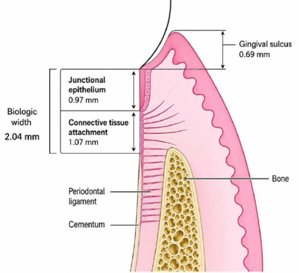
Classical concept of biologic width showing the average dimensions of the junctional epithelium (0.97 mm) and supracrestal connective tissue attachment (1.07 mm), with a combined average biologic width of 2.04 mm. Adapted from Newman MG, Takei HH, Klokkevold PR, Carranza FA. Newman and Carranza’s Clinical Periodontology. 13th ed. Elsevier; 2018.
Although the anatomical components of the supracrestal tissues have long been recognized, clinical interpretation has predominantly focused on their vertical dimension for restorative and periodontal decision-making. This emphasis may not fully reflect the circumferential spatial organization and protective function of these tissues. Therefore, there is a need for a complementary conceptual framework that illustrates the supracrestal tissue attachment as a continuous three-dimensional protective complex while preserving the established biologic width and STA concepts.
Traditional definitions have primarily emphasized the vertical dimension of the supracrestal tissues for diagnostic and restorative purposes. Although this dimensional approach has substantial clinical value, it may not fully emphasize the circumferential organization and three-dimensional protective architecture of the supracrestal tissue attachment. In both natural teeth and dental implants, the junctional epithelium and connective tissue exhibit a continuous circumferential arrangement that extends beyond a simple linear measurement. Therefore, while the concept of biologic width has historically provided an essential dimensional reference, it may benefit from a complementary three-dimensional conceptual framework that better illustrates the spatial organization and protective function of the supracrestal tissues.
It should be emphasized that the proposed biologic belt is not intended to replace the established concepts of biologic width or supracrestal tissue attachment (STA). Rather, it complements these concepts by providing a conceptual three-dimensional visualization of the supracrestal tissue attachment, emphasizing its continuous circumferential organization and protective biological function while preserving the established dimensional interpretation used in clinical practice.
This article proposes a novel conceptual framework, termed the biologic belt, that complements the existing concept of supracrestal tissue attachment (STA) by emphasizing its continuous, circumferential, and three-dimensional protective organization. Rather than introducing a new anatomical structure, the biologic belt provides a conceptual visualization of the supracrestal tissues that may facilitate a more comprehensive understanding of periodontal–restorative relationships around both natural teeth and dental implants. This framework may assist clinical decision-making in restorative margin placement, periodontal procedures, and peri-implant tissue management.
1.1. Proposed Concept: Biologic Belt
1.1.1. Definition
The biologic belt is proposed as a conceptual three-dimensional representation of the supracrestal tissue attachment (STA). It is defined as a continuous, circumferential supracrestal tissue complex surrounding the natural tooth or dental implant, consisting of the junctional epithelium and supracrestal connective tissue. Collectively, these tissues function as a protective biological barrier against mechanical, microbial, and restorative challenges while maintaining the integrity of the underlying periodontal and peri-implant tissues (Fig. 2).
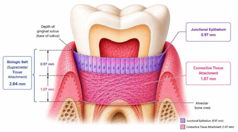
Proposed concept of the biologic belt. Unlike the traditional linear interpretation of biologic width, the biologic belt illustrates the supracrestal tissue attachment as a continuous three-dimensional circumferential structure extending from the base of the gingival sulcus to the alveolar bone crest. It comprises the junctional epithelium (0.97 mm) and the supracrestal connective tissue attachment (1.07 mm), with a total average vertical dimension of 2.04 mm.
1.2. Structural Components
The biologic belt is composed of three principal structural elements that collectively establish its protective function and three-dimensional organization. Rather than representing isolated anatomical structures, these components act together to form a continuous supracrestal tissue complex surrounding the tooth or implant. (Table 1).
| Component | Location | Principal Function |
|---|---|---|
| Junctional Epithelium (JE) | Apical to the gingival sulcus | Provides epithelial attachment and forms a biological seal against microbial invasion |
| Supracrestal Connective Tissue (CT) | Coronal to the alveolar crest, surrounding the tooth or implant | Provides mechanical support, connective tissue attachment, and contributes to the protective soft tissue barrier |
| Circumferential Fiber Apparatus | Dentogingival, circular, transseptal, and supracrestal collagen fibers | Maintains circumferential tissue integrity, stabilizes the soft tissue complex, and distributes functional forces |
1.3. Comparison with Biologic Width
Biologic width has traditionally been described as the vertical dimension of the supracrestal tissues, extending from the alveolar bone crest to the base of the gingival sulcus and comprising the junctional epithelium and supracrestal connective tissue attachment [2]. This dimensional concept remains fundamental for restorative and periodontal treatment planning.
The proposed biologic belt does not replace this established concept. Rather, it complements biologic width by providing a three-dimensional conceptual framework that emphasizes the circumferential continuity, spatial organization, and protective biological function of the supracrestal tissues. While biologic width is a linear clinical measurement, the biologic belt provides a conceptual visualization of the same tissue complex from a three-dimensional perspective, facilitating a more comprehensive understanding of periodontal and peri-implant tissue architecture.
1.4. Natural Teeth
The biologic belt concept provides a three-dimensional perspective for restorative and periodontal treatment planning by emphasizing the preservation of the circumferential supracrestal tissue architecture. Clinically, this framework may facilitate biologically appropriate crown margin placement, periodontal surgical procedures, crown lengthening, and management of deep subgingival restorations, thereby reducing the risk of disrupting the protective soft-tissue complex.
1.5. Dental Implants
A comparable biologic belt may also be considered around dental implants; however, its structural organization differs from that of natural teeth because of the absence of the periodontal ligament and the distinct orientation of peri-implant connective tissue fibers. In addition, peri-implant supracrestal tissue dimensions are generally greater than those around natural teeth, with important implications for implant positioning, emergence profile design, and peri-implant soft tissue management [8, 9].
2. DISCUSSION
The concept of biologic width has served as a fundamental principle in restorative dentistry and periodontology for several decades, providing an important dimensional guideline for maintaining periodontal health. However, in clinical practice, its application has predominantly emphasized the vertical dimension of the supracrestal tissues. Although this approach has considerable diagnostic and therapeutic value, it may not fully illustrate the circumferential organization and three-dimensional protective architecture of the supracrestal tissue attachment.
The biologic belt is proposed as a conceptual framework that complements, rather than replaces, the established concepts of biologic width and supracrestal tissue attachment (STA). Rather than introducing a new anatomical structure, it provides a three-dimensional conceptual visualization of the same supracrestal tissue complex by emphasizing its continuous circumferential organization and protective biological function. This perspective is consistent with the contemporary understanding of STA as a composite epithelial and connective tissue attachment that serves as an essential biological barrier protecting the underlying periodontal tissues [10, 11].
From a clinical perspective, the biologic belt concept may facilitate a more comprehensive interpretation of restorative and periodontal procedures by encouraging clinicians to consider not only the vertical dimension but also the circumferential integrity of the supracrestal tissues. This perspective may be particularly relevant during restorative margin placement, crown lengthening procedures, management of deep subgingival restorations, and peri-implant soft tissue management.
A comparable biologic belt may also be considered around dental implants. Although the peri-implant soft tissue performs a similar protective function, its structural organization differs from that surrounding natural teeth because of the absence of the periodontal ligament and differences in connective tissue fiber orientation. Furthermore, peri-implant supracrestal tissue dimensions are generally greater than those around natural teeth, which may influence peri-implant health, esthetics, and restorative planning [6].
The present article introduces a conceptual framework rather than quantitative anatomical measurements. Consequently, the proposed biologic belt should not be interpreted as a new measurable anatomical entity. Future investigations employing histological analysis, micro-computed tomography, cone-beam computed tomography, digital three-dimensional reconstruction, and prospective clinical studies are required to validate this concept and establish reproducible dimensional parameters.
Overall, the biologic belt should be regarded as a conceptual extension of biologic width and STA, providing an additional three-dimensional perspective for understanding the organization and protective function of the supracrestal tissues while preserving the established dimensional principles that remain fundamental to periodontal and restorative treatment planning.
CONCLUSION
The biologic belt is proposed as a conceptual three-dimensional framework that complements the established concepts of biologic width and supracrestal tissue attachment (STA) by emphasizing the continuous circumferential organization and protective function of the supracrestal tissues. Rather than replacing these well-established concepts, the biologic belt provides an additional three-dimensional perspective that may facilitate a more comprehensive understanding of periodontal–restorative relationships around both natural teeth and dental implants. If validated by future experimental and clinical studies, this conceptual framework may contribute to biologically guided restorative and periodontal treatment planning.
RECOMMENDATION
Future research should focus on validating the biologic belt concept through histological investigations, cone-beam computed tomography (CBCT), micro-computed tomography (micro-CT), digital three-dimensional reconstruction, finite element analysis, and well-designed prospective clinical studies. These investigations should establish reproducible measurements of the vertical dimension, buccolingual and mesiodistal tissue thickness, and circumferential continuity of the supracrestal tissue complex in both natural teeth and dental implants.
Further experimental and histological studies are also required to determine whether the proposed biologic belt provides clinically relevant information beyond the established dimensional assessment of biologic width and supracrestal tissue attachment. Validation of this conceptual framework may contribute to improved biologically based restorative, periodontal, and peri-implant treatment planning.
AUTHORS' CONTRIBUTIONS
The authors confirm their contributions to the paper as follows: O.E.: Presenting the original idea and concept creation. Writing the article, article submission, and writing article; G.T.: Writing, translation, and editing the article, assisting in article submission.
LIST OF ABBREVIATIONS
| JE | = Junctional Epithelium |
| STA | = Supracrestal Tissue Attachment |
| CBCT | = Cone-beam Computed Tomography |
ACKNOWLEDGEMENTS
Declared none.