All published articles of this journal are available on ScienceDirect.

Cytotoxic Effects of Ethyl Acetate and Hexane Extracts of Capsicum annuum L. on Cal 27 Oral Cancer Cells

Abstract
Introduction
This study aims to compare the cytotoxic effects of ethyl acetate and n-hexane extracts of Capsicum annuum L. on Cal27 oral cancer cell lines by examining caspase-3 expression.
Materials and Methods
This study used an in vitro experimental design with a post-test-only control group. Capsaicin in the ethyl acetate extract of Capsicum annuum was identified using Thin Layer Chromatography (TLC) with detection at UV 254 nm. Cytotoxicity was evaluated using the MTT assay, morphological assessment, and caspase-3 expression. Caspase-3 expression was quantified using qPCR with GAPDH, and relative expression was calculated by the 2^–ΔΔCt method. Statistical Analysis: Differences in caspase-3 expression between selected treatment groups were examined using paired two-sample t-tests, whereas overall variation among all groups was analyzed with one-way ANOVA. Bonferroni post hoc tests were applied in multiple comparisons. Statistical significance was defined as p < 0.05 and adjusted to p < 0.005 for multiple testing.
Results
Phytochemical testing confirmed the presence of capsaicin in the extract. TLC analysis on silica gel GF 254 using a toluene–ethyl acetate–formic acid (5:4:1 v/v/v) mobile phase showed a single blue-violet spot (Rf = 0.71) under UV 254 nm, consistent with the capsaicin standard. Both fractions reduced the viability of Cal 27 cells in a concentration-dependent manner. The n-hexane fraction showed stronger cytotoxic activity, with an IC50 of 218.10 µg/mL, compared with 317.10 µg/mL for the ethyl acetate fraction. Morphological examination revealed features consistent with apoptosis, such as apoptotic body formation. Under certain conditions (medium-only, 2% DMSO, and 1 × IC50), caspase-3 expression appeared slightly higher in the ethyl acetate fraction; however, these differences were not statistically significant (p > 0.05). Although ANOVA indicated overall significance within the hexane-treated group (p = 0.005), Bonferroni-corrected post hoc analysis did not find any significant pairwise differences.
Discussion
The stronger cytotoxic effect observed with the n-hexane fraction suggests that non-polar constituents may play a major role in suppressing Cal 27 cell proliferation. While both extracts produced morphological evidence of apoptosis, the lack of significant variation in caspase-3 expression shows that additional mechanisms may be involved. Variations in solvent polarity likely influenced the phytochemical profiles of the extracts and their biological activity.
Conclusion
Both n-hexane and ethyl acetate fractions of C. annuum exhibited cytotoxic effects on Cal 27 oral cancer cells and produced morphological changes consistent with apoptotic features; however, further studies are required to confirm the involvement of apoptotic pathways.
1. INTRODUCTION
Cancer causes approximately 9.7 million deaths annually worldwide, with an estimated one in nine men and one in twelve women dying from the disease [1, 2]. Oral cancer is the 16th most common cancer globally [3]. Tongue cancer is the most prevalent oral cancer [4, 5], which is characterized by aggressive local invasion and a high susceptibility to metastasis to cervical lymph nodes [6]. The survival rates of patients with tongue cancer are quite low due to disease recurrence driven by local invasion and metastasis [7]. Given its poor prognosis, early diagnosis is crucial for improving treatment outcomes [8].
Cancers were commonly treated using surgery, radio-therapy, and chemotherapy [9]. However, those treat-ments require high cost and cause significant financial, psychological, and social burdens on both patients and healthcare systems [10]. Furthermore, these treatments are frequently associated with long-term adverse effects, including fatigue, anorexia, diarrhea, sleep disturbances, hot flashes, neuropathy, and chronic pain [11, 12], which can affect the patients’ quality of life [10].
Therefore, research on alternative or supportive cancer therapies that have minimal side effects has gained increasing attention, such as the use of natural products or herbal medicines. The bioactive compounds, which are the secondary metabolites of the herbal medicine, are the ones that contribute to their pharmacological activities [13]. One example is capsaicin, the main active compound found in Capsicum annuum L [14]. This plant is known for its wide range of biological activities, including anticancer activity against oral cancer [15]. Previous experimental studies reported that capsaicin can inhibit the proliferation of oral cancer cells, induce apoptosis, and suppress tumor initiation and progression in both in vitro and in vivo models [16-20].
The efficiency of extracting bioactive compounds depends on the choice of the solvent used [21]. Choosing the right solvent must consider factors such as selectivity, solubility, cost, and safety [22]. N-hexane is often used as a non-polar solvent because it effectively extracts non-polar compounds, has a low boiling point that makes evaporation and recovery easier, and has low viscosity that enhances penetration and extraction efficiency [23]. On the contrary, ethyl acetate has moderate polarity, allowing it to extract both polar and non-polar compounds. It also has strong dissolving ability for bioactive molecules such as flavonoids, phenolic acids, and terpenes, and is considered relatively safe for pharmaceutical use [24]. Based on these considerations, this study aims to compare the cytotoxic effects of ethyl acetate and n-hexane extracts of Capsicum annuum L. on Cal27 oral cancer cell lines by examining caspase-3 expression.
2. MATERIALS AND METHODS
2.1. Study Design
This study was an in vitro experimental design using a post-test-only control group. Cytotoxicity was evaluated using the MTT assay, morphological assessment, and caspase-3 expression.
2.2. Materials and Reagents
The materials used in this study included hexane and ethyl acetate fractions of Capsicum annuum L. Capsaicin in the ethanol extract of Capsicum annuum was identified using Thin Layer Chromatography (TLC). Cal 27 oral squamous carcinoma cells (ATCC CRL-2095), Dulbecco’s Modified Eagle Medium (DMEM) supplemented with 10% Fetal Bovine Serum (FBS), 1% streptomycin-penicillin, and 1% fungizone, MTT reagent (3-(4,5-dimethylthiazol-2-yl)-2,5-diphenyl tetrazolium bromide), PrestoBlue™, cisplatin as a positive control, 2% dimethyl sulfoxide (DMSO) as a vehicle control, DEPC-treated water, RNA extraction reagent (GENEzol™), and reagents for reverse transcription PCR.
2.2.1. Extraction of Capsicum annuum L
Fresh red chilli (Capsicum annuum L.) was obtained from Pangalengan, Bandung Regency, West Java, Indonesia. A total of 5 kilograms of chilli was washed, drained, and thinly sliced before being dried in the shade. The dried material was ground into a fine powder and sieved through a 1 mm mesh. The resulting powder was macerated in ethanol at 37°C for three consecutive 24-hour periods with agitation every four hours. The extract was filtered daily, and the combined filtrates were concentrated using a rotary vacuum evaporator to obtain a thick ethanol extract. Ten percent of this extract was stored. The remaining 90% of the extract underwent liquid-liquid partitioning using hexane, dichloromethane, and ethyl acetate. The hexane and ethyl acetate fractions were collected and evaporated prior to cytotoxicity testing.
2.2.2. Thin-layer Chromatography (TLC) Analysis
TLC analysis was performed using silica gel 60 GF254 plates with a mobile phase consisting of toluene–ethyl acetate–formic acid (5:4:1 v/v/v). The Capsicum annuum extract and standard capsaicin were spotted onto the plate and eluted to a distance of 8 cm. The spots were visualized under UV light at 254 nm after derivatization with vanillin–sulfuric acid reagent.
2.2.3. Cal 27 Cell Culture
Cal 27 cells were obtained from the Central Laboratory of Universitas Padjadjaran. The cells were cultured in DMEM containing 10% FBS, 1% streptomycin-penicillin, and 1% fungizone. The cells were maintained in a humidified incubator at 37°C with 5% CO2. Cells were subcultured at 70–80% confluency using 0.25% trypsin-EDTA.
2.2.4. Cytotoxicity Assay
The cytotoxic effects of hexane and ethyl acetate extracts were evaluated using the MTT assay. Cal 27 cells were seeded into 96-well plates at a density of 5 × 10³ cells per well and incubated overnight. The following day, the cells were treated with four serial concentrations of each extract (125, 250, 500, and 1000 µg/mL) and incubated for 24 hours. Each treatment group, including controls, was replicated four times (n = 4). The control groups consisted of cisplatin (positive control), 2% DMSO (vehicle control), and a medium-only group with untreated cells. After 24 hours of treatment, 10 µL of MTT reagent was added to each well and incubated for 4 hours. Formazan crystals formed were dissolved using PrestoBlue™ reagent, and absorbance was measured at 595 nm using a microplate reader.
2.2.5. IC50 Determination and Treatment Design
IC50 values for each extract were calculated by plotting log concentrations against cell viability followed by linear regression analysis. These IC50 values were used to define the concentrations for subsequent experiments, specifically at 1 × IC50 and 2 × IC50. These doses, together with positive and negative controls, were applied to assess morphological changes and gene expression levels of caspase-3.
2.2.6. Morphological Observation
Morphological changes in Cal 27 cells were observed to evaluate the effects of the treatments. Cells were seeded at a density of 1 × 10⁶ in 24-well plates and cultured until approximately 80% confluence. The cells were then treated with hexane and ethyl acetate extracts at 1 × and 2 × IC50 doses, cisplatin, and 2% DMSO for 24 hours. Changes in cell morphology were observed under a light microscope and documented.
2.2.7. RNA Extraction
RNA extraction was performed using the TRIzol-based GENEzol™ method. After the treatment, the adherent cells were washed with cold Phosphate-Buffered Saline (PBS) and lysed with 200 µL GENEzol™ reagent per well. The lysates were transferred to microcentrifuge tubes, and 40 µL of chloroform was added. After vigorous mixing for 15 seconds, the samples were incubated at room temperature for 3 minutes and centrifuged at 11,200 rpm for 15 minutes. The aqueous phase was transferred to a new tube and mixed with 100 µL of isopropanol. After a 10-minute incubation, the samples were centrifuged again to pellet the RNA. The pellet was washed with 70% ethanol, centrifuged, air-dried, and dissolved in 20 µL of DEPC-treated water. The RNA was incubated at 60°C for 10 minutes and stored at −70°C for further analysis.
2.2.8. Quantitative PCR (qPCR)
qPCR was performed to evaluate the mRNA expression level of caspase-3 in Cal 27 cells using a one-step qPCR kit. The 10 µL reaction mixture consisted of 2 µL of RNA template, 0.4 µL of forward primer, 0.4 µL of reverse primer (each at 10 µM), 5 µL of 2 × SensiFAST SYBR Green mix, 0.1 µL of reverse transcriptase, 0.2 µL of RNase inhibitor, and 1.9 µL of DEPC-treated water. The thermal cycling conditions included reverse transcription at 45°C for 10 minutes, an initial denaturation at 95°C for 2 minutes, followed by 40 amplification cycles of denaturation at 95°C for 5 seconds and annealing-extension at 60–65°C for 20 seconds. A melting curve analysis was performed at the end to confirm amplification specificity. The housekeeping gene glyceraldehyde-3-phosphate dehydrogenase (GAPDH) was used as an internal control to normalize the expression levels of caspase-3, and relative expression was calculated using the 2^–ΔΔCt method. Each experiment was performed using four biological replicates (n = 4), with each measurement conducted in technical duplicate.
2.3. Statistical Analysis
Data normality and homogeneity of variances were assessed using boxplots and the Shapiro-Wilk test. Paired two-sample t-tests were used to determine the differences in caspase-3 expression between specific treatment groups. To assess the overall differences in caspase-3 expression among all treatment groups, a one-way Analysis of Variance (ANOVA) was used. If significant differences were detected, a Bonferroni post hoc test was applied. Statistical significance was set at p < 0.05, with an adjusted threshold of p < 0.005 applied for the Bonferroni correction.
3. RESULTS
TLC analysis of the ethanol extract of Capsicum annuum revealed a single blue-violet spot (Rf = 0.71) under UV light at 254 nm after derivatization with vanillin–sulfuric acid reagent, consistent with the capsaicin standard. The results showed that all tested concentrations of Capsicum annuum extracts inhibited the growth of Cal 27 cells. The IC50 value of the n-hexane extract was 218.10 µg/mL, while the ethyl acetate extract had an IC50 of 317.10 µg/mL. After 24 hours of incubation, treated cells showed morphological changes compared with those in the negative control group (Fig. 1). Cells exposed to both hexane and ethyl acetate extracts at different concentrations exhibited typical apoptotic features, including the formation of apoptotic bodies. Similar morphological changes were also observed in the cisplatin-treated positive control group. These apoptotic bodies appeared as small, round structures measuring approximately 1–5 μm in diameter.
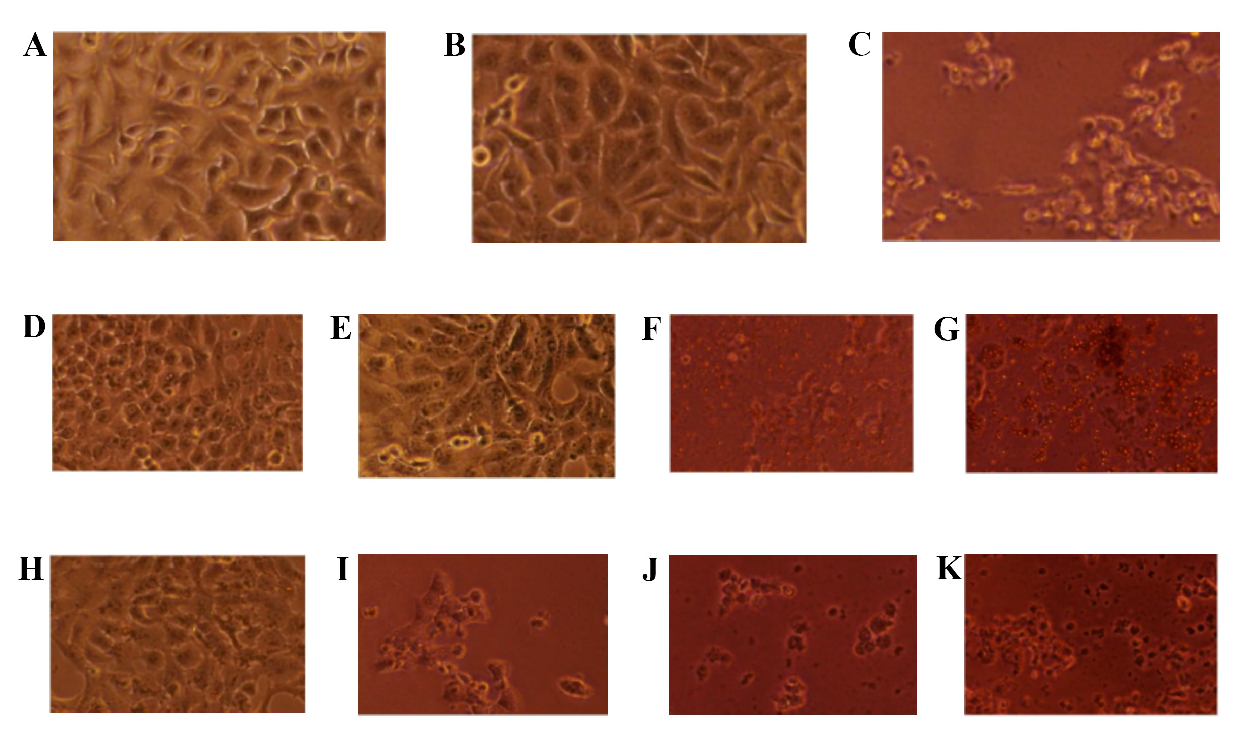
Representative micrographs of Cal 27 cells after 24-hour of incubation under different treatments: medium-only control (A), 2% DMSO (B), cisplatin (C), ethyl acetate extract at 125 µg/mL (D), 250 µg/mL (E), 500 µg/mL (F), and 1000 µg/mL (G), and hexane extract at 125 µg/mL (H), 250 µg/mL (I), 500 µg/mL (J), and 1000 µg/mL (K). DMSO, dimethyl sulfoxide.
The relative expression of caspase-3 was compared among different treatment groups for both ethyl acetate and hexane fractions of Capsicum annuum (Table 1). A one-way ANOVA revealed no significant differences among the groups treated with the ethyl acetate fraction (p = 0.062). For the hexane fraction, ANOVA indicated an overall significant difference among groups (p = 0.005). However, subsequent Bonferroni post hoc analysis, with an adjusted significance threshold of p < 0.005, showed that none of the pairwise comparisons were statistically significant.
| Treatment | Groups | Mean | SEM | p-value |
|---|---|---|---|---|
| Ethyl acetate | Medium | 1.01 | 0.15 | 0.062 |
| Vehicle control (2% DMSO) | 1.43 | 0.18 | ||
| Positive control (Cisplatin) | 1.92 | 0.30 | ||
| 1 x IC50 | 1.40 | 0.08 | ||
| 2 x IC50 | 2.22 | 0.34 | ||
| Hexane | Medium | 0.76 | 0.24 | 0.005* |
| Vehicle control (2% DMSO) | 1.11 | 0.29 | ||
| Positive control (Cisplatin) | 2.26 | 0.45 | ||
| 1 x IC50 | 0.94 | 0.23 | ||
| 2 x IC50 | 2.24 | 0.36 |
Note: *Statistically significant difference according to one-way ANOVA.
Figure 2 shows that in some treatment conditions (medium-only, 2% DMSO, and 1 × IC50), the ethyl acetate fraction group exhibited slightly higher caspase-3 expression than the hexane fraction group. However, paired two-sample t-tests revealed no statistically significant differences between the ethyl acetate and hexane fraction groups for any treatment condition (p > 0.05 for all comparisons).
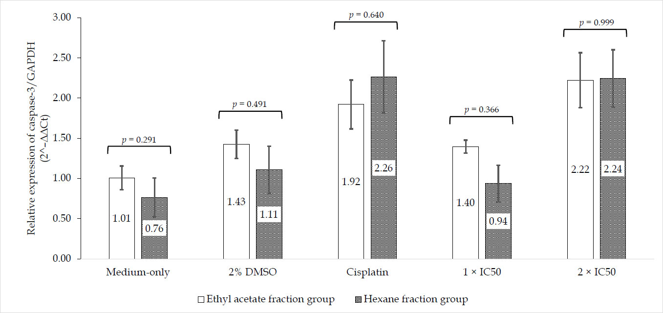
Caspase-3 expression in Cal 27 cells treated with ethyl acetate or hexane fractions of Capsicum annuum L under various conditions. Data represent mean ± SEM (n=4). Statistical differences between corresponding groups were evaluated using paired two-sample t-tests (p < 0.05). DMSO, dimethyl sulfoxide.
4. DISCUSSION
The TLC findings indicated the presence of capsaicin in the ethanol capsicum annum extract, consistent with previous reports showing that capsaicin produces a blue-violet spot with an Rf value of 0.70–0.72. The findings of this study indicate that Capsicum annuum extracts obtained using n-hexane and ethyl acetate both exerted cytotoxic effects on Cal 27 oral cancer cells. The inhibition of cell proliferation occurred in a concentration-dependent manner. The n-hexane fraction (IC50 = 218.10 µg/mL) showed greater cytotoxic activity than the ethyl acetate fraction (IC50 = 317.10 µg/mL), which suggests that the non-polar bioactive compounds extracted by n-hexane may play a major role in the observed anticancer effect. This finding is consistent with previous studies reporting that lipophilic phytochemicals, such as capsaicinoids and carotenoids, are more efficiently extracted using non-polar solvents and may contribute significantly to antiproliferative activity in cancer cells [25, 26].
Morphological observations further supported the cytotoxic results. Cells treated with both extracts exhibited characteristic apoptotic features, including the formation of apoptotic bodies, similar to those seen in cisplatin-treated cells. The presence of apoptotic bodies is a classic morphological sign of programmed cell death and is consistent with previous evidence showing that capsaicin and related compounds in Capsicum annuum can trigger apoptosis through mitochondrial and caspase-dependent pathways [15].
Caspase-3 was evaluated to assess whether apoptosis induction may be associated with caspase-dependent mechanisms. Although the relative expression of caspase-3 was slightly higher in the ethyl acetate fraction than in the hexane fraction, statistical analysis showed no significant differences between groups. The hexane fraction showed overall significance in the ANOVA analysis; however, post hoc comparisons were not statistically significant after adjustment. This discrepancy may be due to the conservative nature of Bonferroni correction and the limited sample size (n = 4), which reduces statistical power [27]. These findings suggest that while both extracts may induce morphological features consistent with apoptosis, caspase-3 activation alone may not fully explain the observed cytotoxic effects [14]. Therefore, the involvement of apoptotic pathways should be interpreted cautiously, as additional apoptosis-related markers, including Annexin V, Bax/Bcl-2, caspase-8, caspase-9, reactive oxygen species generation, and mitochondrial function assays, were not evaluated in the present study [28].
Capsaicin treatment may be associated with apoptosis-related changes in Cal 27 cell lines, as indicated by caspase-3 expression. Caspase-3 is a key executioner caspase involved in the final stage of apoptosis, functioning in both intrinsic and extrinsic pathways [29]. In general, apoptosis occurs through two main mechanisms. The extrinsic pathway is initiated by the interaction between death receptors on the cell membrane and their ligands, leading to activation of initiator caspase-8 [29, 30]. The intrinsic pathway involves mitochondrial signaling, including the release of cytochrome c, which activates initiator caspase-9 [29, 30]. Both pathways converge at the activation of caspase-3 as a downstream effector [30].
Although initiator caspases (caspase-8 and caspase-9) were not examined due to resource limitations, the measurement of caspase-3 provides preliminary insight into apoptosis-related processes [31, 32]. Previous research by Kurnijasanti et al. demonstrated that Capsicum annuum extract inhibited the growth of T47D breast cancer cells and was associated with increased caspase-3 expression, supporting the use of caspase-3 as an apoptosis indicator [33].
Although the ethyl acetate fraction showed slightly higher caspase-3 expression at certain treatment conditions, its overall cytotoxicity was lower than that of the hexane fraction. This discrepancy suggests that different classes of phytochemicals may act via distinct apoptotic or non-apoptotic mechanisms. Ethyl acetate is known to extract semi-polar compounds such as flavonoids and phenolic acids, which can exert anticancer effects via antioxidant modulation, inhibition of angiogenesis, or suppression of key signaling pathways such as NF-κB and PI3K/Akt [26, 34-36]. Meanwhile, hexane-extracted non-polar compounds (e.g., capsaicinoids, carotenoids, and sterols) are often more directly cytotoxic and pro-apoptotic in nature [26, 37, 38]. These results suggest that Capsicum annuum extracts, regardless of solvent polarity, possess anticancer potential against oral cancer cells. However, the differences in IC50 values, caspase-3 expression, and phytochemical content highlight the complexity of plant-derived anticancer activity.
This study has several limitations. It was conducted in vitro using a single oral cancer cell line, which may not reflect overall tumor behavior in a living organism. Phytochemical analysis was limited to preliminary identification using TLC, which indicated the presence of capsaicin. Therefore, the specific compounds responsible for the observed cytotoxic activity could not be definitively identified or quantified. Differences in research results can be influenced by variations in testing methods, cancer cell culture conditions, and the characteristics of the extracts used in each study. Variations in bioactive compounds in the extract are also influenced by locations of the plant’s growth, harvest age, and extraction method. These factors can affect the anticancer activity obtained. In addition, the cytotoxic mechanism was only assessed through caspase-3 expression, so other apoptotic or non-apoptotic pathways may have been overlooked. The relatively small number of replicates may also have limited statistical power. Further studies using other mechanistic markers, detailed phytochemical profiling, and in vivo models are needed to confirm and extend these findings.
CONCLUSION
This study demonstrated that Capsicum annuum extracts obtained using n-hexane and ethyl acetate exert cytotoxic effects on Cal 27 oral cancer cells. Both fractions produced morphological changes consistent with apoptotic features, similar to those observed with cisplatin, although caspase-3 expression did not differ significantly between the two groups. However, the involvement of apoptotic pathways requires further confirmation through additional assays.
AUTHORS’ CONTRIBUTIONS
The authors confirm contribution to the paper as follows: W.Y.: Study conception and design; W.Y.: Data collection; W.Y., A.R.: Analysis and interpretation of results; A.R.: Draft manuscript. All authors reviewed the results and approved the final version of the manuscript.
LIST OF ABBREVIATIONS
| TLC | = Thin Layer Chromatography |
| UV | = Ultra Violet |
| ANOVA | = Analysis of Variance |
AVAILABILITY OF DATA AND MATERIALS
The data and supportive information are available within the article.
ACKNOWLEDGEMENTS
Declared none.