All published articles of this journal are available on ScienceDirect.

Uncommon Inferior Alveolar Nerve Injury Associated with the Mandibular Second Molar: A Case Report
 iD
iD
Authors Info & Affiliations
Abstract
Introduction
Inferior alveolar nerve injury is most frequently associated with mandibular third molar surgery, while neurosensory complications related to endodontic treatment are uncommon and sparsely reported. Such injuries may occur when mandibular molar roots and associated periapical lesions are located in proximity to the inferior alveolar canal, allowing extruded endodontic materials to affect the nerve. This case adds to the limited scientific literature by describing delayed-onset inferior alveolar nerve sensory impairment following root canal treatment and its recovery after elimination of the causative factor.
Case Presentation
A 50-year-old woman presented with numbness of the right lower lip and chin that developed three months after root canal treatment of the mandibular right second molar. Clinical examination and radiographic evaluation using an orthopantomogram and cone beam computed tomography demonstrated extruded gutta-percha extending into the inferior alveolar nerve canal, associated periapical radiolucency, and direct contact between the tooth roots and the nerve. The mandibular right first molar also showed residual roots with periapical pathology. Following informed consent, atraumatic extraction of both teeth was performed, followed by socket debridement and supportive management including vitamin B12 supplementation. At four weeks, neurosensory examination showed persistent anesthesia. Gradual improvement was noted at eight weeks, and near complete recovery of sensation was observed at twelve weeks after extraction.
Conclusion
This case demonstrates that inferior alveolar nerve injury can occur as a rare complication of endodontic treatment of mandibular molars, particularly when extruded filling materials and periapical pathology are present close to the nerve canal. Early recognition, careful radiographic assessment, and timely removal of the etiologic factor may facilitate favorable neurosensory recovery and reduce the risk of permanent sensory deficits.
1. INTRODUCTION
The Inferior Alveolar Nerve (IAN), a branch of the mandibular nerve, travels bilaterally within the mandibular canal, which houses the neurovascular bundle and provides somatosensory sensation to the mandibular teeth. It follows an oblique downward and forward path through the mandibular ramus and curves horizontally near the alveolar region housing the tooth sockets. It runs from the mandibular foramen on the lingual side of the mandibular ramus to the mental foramen on the buccal surface of the mandibular body [1]. Moreover, IAN provides motor innervation to the mylohyoid muscle and sensory innervation to all mandibular teeth, the gingiva, the lower lip, and the chin while it transmits pain signals from these areas and gives rise to the mental nerve, which supplies the skin and mucous membrane of the mandibular incisor area [2].
Complications involving the IAN can arise from various etiological factors, including pathological conditions, mechanical trauma, and surgical interventions such as mandibular third molar extractions and endodontic treatment [3]. Although less common with root canal treatment, endodontic treatment can also be responsible for neurosensory injuries, which can result in unfavorable anesthesia, paresthesia, and dysesthesia [3, 4].
Eight percent of inferior alveolar nerve damage is associated with endodontic treatment [5].
The proximity of the mandibular molars' apices to the IAN was measured as less than 1 mm from the apices to the mandibular canal, which contained the neurovascular bundle. This close distance means that the extrusion of endodontic materials may cause chemical, mechanical, or heat-related damage to the nerve by breaching the mandibular canal [6]. Moreover, damage to the IAN is influenced by its position relative to the tooth roots. The IAN is found to be in a lingual position in 35.4% of cases, in a buccal position in 30.8%, in an apical position in 27%, and interradicular in 6.9%, with neurosensory impairments more frequently observed when the IAN is positioned directly lingual to the tooth roots [7]. Moreover, El Hadidi et al. conducted a Cone Beam Computed Tomography (CBCT) analysis of 170 cases to assess the relationship between mandibular teeth and the inferior alveolar nerve. The findings revealed that the foramen was positioned behind the second premolar in 79% of cases, whereas in only 21%, the nerve extended to the first premolar region. In the premolar area, the presence of a loop influenced the gap between the root apex and the uppermost point of the superior cortex of the inferior alveolar nerve canal [8].
In a case report by Kasapoğlu and Doğancalı (2020), an overflow of calcium hydroxide from the apical part of the root into the inferior alveolar nerve canal was documented, causing injury, particularly postoperative paresthesia. Owing to the nature of calcium hydroxide and its short resorption time, the material was completely resorbed after 6 months, and the degree of paresthesia decreased. It disappeared completely at the 3-year follow-up visit [5]. Another case report by DĠNÇER et al. (2021) presented the case of a 40-year-old woman who experienced IAN paresthesia in her right lower lip and chin caused by overextension of the gutta-percha two days after endodontic treatment. To address the patient’s complaint, the overextended gutta-percha was removed from the root canal, and the her symptoms were resolved [9]. Additionally, in a 2001 case report by Amailuk et al., an unexpected neurosensory deficit following tooth extraction of a mandibular first molar was attributed to possible causes, including trauma from the dental injection, local infection, or pressure on an accessory branch during the procedure. This highlights the multifactorial nature of neurologic deficits, even in routine dental extractions [10]. With respect to recovery after injury to the IAN, postoperative recovery occurred most notably at 3 months for the IAN and at 6 months for the LN, with complete recovery achieved in 67% of IAN deficits and 72% of LN deficits by the end of follow-up [11]. A study conducted in 2004 by Queral-Godoy et al. to assess the incidence of inferior alveolar nerve lesions after lower third molar extractions revealed that most cases of IAN impairment resolve within six months, although recovery can extend beyond one year, with older patients facing a greater risk of incomplete restoration of chin and lip sensation [12].
While most cases of IAN injury are associated with third molar extractions, injuries following root canal treatment, particularly involving the mandibular second molar, are rare and only a limited number of cases have been reported in the literature. Consequently, evidence regarding their diagnosis, management, and prognosis remains scarce. The present case adds to the limited evidence available. A better understanding of the mechanisms, clinical presentation, and recovery patterns of IAN injury following endodontic therapy may help guide future research and improve prevention and treatment approaches.
2. CASE PRESENTATION
A 50-year-old woman was referred to the Oral and Maxillofacial Surgery Department at King Abdullah Medical City Specialist Hospital in Makkah, Saudi Arabia, for extraction of the mandibular right first molar (MR6) and mandibular right second molars (MR7). She was initially evaluated in the emergency department, where she presented with complaints of numbness in the lower right region of her jaw. She had undergone root canal treatment on MR7, and the numbness started three months later, at which time she presented to our clinic, as the lesion expanded due to an extruded gutta-percha, persisting until the day of evaluation
2.1. Investigations
Upon clinical and radiographic examination, in the area of interest, MR7 was found to be restored with recurrent caries and had root canal treatment, and was tender to percussion. The remaining MR6 root that was indicated for extraction had prior endodontic treatment. An orthopantomogram (OPG) radiograph (Fig. 1) and CBCT (Fig. 2) of MR7 demonstrated a periapical round radiolucency measuring approximately 0.5 millimeters in diameter around both the mesial and distal roots, with extruded gutta-percha. Additionally, the roots were in contact with the IAN. For MR6, the remaining roots showed periapical pathology. A detailed review of the patient’s medical history revealed no significant findings, with no history of systemic diseases, chronic illnesses, diabetes, hypertension, cardiovascular disorders, or other notable medical conditions.
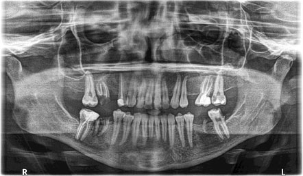
Preoperative OPG revealed the presence of extruded gutta-percha within the area of the mandibular canal.
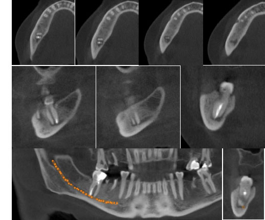
Preoperative CBCT with different views revealed the presence of extruded gutta-percha percha and periapical lesions within the IAN.
2.2. Management
After informing the patient about the treatment options and possible complications, the patient decided to proceed with the extraction. Signed consent was obtained, and the patient was prepared for the procedure under local anesthesia. A right IAN block, long buccal nerve block, and LN block were administered using 2% lidocaine with 1:100,000 epinephrine, with 1.8 mL injected for the IAN and LN blocks and 0.5 mL for the long buccal nerve block, ensuring negative blood aspiration prior to administration.
The extraction of MR7 and MR6 was performed using a combination of conventional forceps and an elevator approach without raising a mucoperiosteal flap. Initial luxation was achieved via a couplant elevator with slow and steady force, followed by the use of lower molar forceps to complete the extraction (Fig. 3).
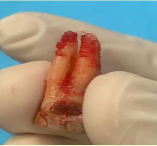
The extracted MR7 with extruded gutta-percha.
The procedure was conducted with minimal trauma to preserve the integrity of the bony socket and minimize the risk of IAN injury. An immediate OPG was taken post-extraction (Fig. 4).
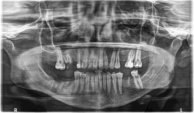
Postoperative OPG after extraction of MR7 and MR6.
Following extraction, soft-tissue remnants were meticulously debrided from the socket with curettes, and the site was irrigated with 40 mL of sterile 0.9% sodium chloride solution. An oxidized cellulose hemostat (SURGICEL® FIBRILLAR) was placed in the socket for hemostasis, and the wound was closed with simple interrupted sutures using a 3-0 chromic gut. Vitamin B12 was prescribed at 500 µg three times daily for 12 weeks to enhance neurosensory recovery. Ibuprofen 400mg was prescribed orally 6 to 8 hours as needed for postoperative pain for 3 days. Postoperative instructions were provided, and the patient was discharged in stable condition.
2.3. Outcome and Follow-up
Two weeks post-extraction, the patient reported that the numbness in the right lip and chin had not changed. Examination revealed an area of numbness over the right lower lip and chin, with no response to touch, pressure, or two-point discrimination, as mentioned in the practical application of clinical neurosensory testing [13].
Four weeks post-extraction, the patient reported no improvement in the area of numbness. Extraoral examination revealed complete anesthesia in the right lower lip and chin region, whereas intraoral examination revealed anesthesia in the mucosa buccal to the right anterior teeth, with paresthesia in the tongue, lingual mucosa, and buccal mucosa of the molar region. Follow-up visits were scheduled once a week after the fourth week.
By the eighth week, as the lesion began to resolve following the removal of the extruded gutta-percha and the debridement of the extraction socket, the patient reported a sign of improvement, indicating that the numbness was reduced in the external corner of the lip and chin, although it remained limited to the buccal mucosa to the right anterior to MR7. Twelve weeks postextraction, both extraoral and intraoral examinations revealed almost complete recovery in the area of anesthesia, with sensitivity to light touch, dull pressure, and two-point discrimination noted.
3. DISCUSSION
The proximity of the IAN to mandibular teeth showed that the closest root apices were those of the second molars at 1.76 mm, followed by the second premolars at 3.09 mm, while in relation to the inferior border of the mandible, the IAN was nearest at the level of the second molar roots at 8 mm, followed by the first molars at 8.1 mm [8].
Nerve injury following endodontic treatment can occur due to various factors, including extrusion of the gutta-percha or endodontic sealer beyond the apical foramen, particularly in teeth located near the IAN, as well as traumatic mechanical or chemical endodontic instrumentation, and these events may lead to serious complications such as permanent nerve damage, extension of infection, bone necrosis, or, in severe cases, mandibular resection [14].
The diagnosis of such cases relies primarily on the reported symptoms and the patient’s medical history, both of which are considered essential. A careful radiographic examination using OPG and CBCT is also recommended owing to the complex anatomy of the nerve and the potential risk of irreversible paresthesia. If nerve injury occurs, an accurate clinical assessment is needed to determine the extent of involvement and to monitor improvement. This requires the thorough application of various techniques, including two-point discrimination, brush directional stroke, and pinprick testing [15, 16].
Extensive periapical lesions increase the risk of IAN injury by compromising the osseous barrier that separates the tooth apex from the neurovascular bundle, therefore allowing the diffusion of neurotoxic byproducts, irrigation, and root canal filling materials during treatment [17].
A case report by Giuliani et al. (2001) described a patient who presented with a history of numbness in the lip, chin, and buccal mucosa following endodontic treatment of MR7, which was in proximity to the IAN. After unsuccessful nonsurgical endodontic retreatment, the tooth is extracted, leaving residual numbness confined to the distribution of the mental nerve [18]. This finding suggests that in cases with a periapical lesion adjacent to the IAN and extrusion of endodontic material, extraction may be preferable to nonsurgical retreatment to minimize the risk of additional nerve injury resulting from further apical extrusion of filling materials, mechanical instrumentation during root canal debridement, or extrusion of endodontic irrigants. In addition to mechanical compression, extruded AH Plus sealer may cause chemical injury through the release of formaldehyde and unreacted epoxy resin components during polymerization, resulting in local inflammation and neural irritation. This highlights that both the chemical composition and the location of the extruded material influence the severity of nerve injury [4, 14].
Additionally, a similar study by Ali et al. (2024) with a four-year follow-up period reported on a patient with lower lip paresthesia due to extruded intracanal medicament in the IAN canal related to MR7. Initial management with canal irrigation was ineffective, but the use of steroids and pregabalin as adjuncts to nonsurgical root canal treatment ultimately led to complete resolution of the symptoms [19]. Moreover, Buyukkurt et al. (2011) reported a case of lower lip paresthesia caused by extruded zinc oxide eugenol on MR7, in which the clinician decided to perform surgical management to remove the extruded material, resulting in complete resolution of the patient’s symptoms within four months [20].
According to a 2016 systematic review by Rosen et al., the prognosis of nerve injury due to extruded endodontic material is influenced by several factors, including tooth location, type of extruded material, obturation technique, type of treatment, and timing of intervention. To improve the prognosis, careful analysis of the anatomy and nature of the problem preoperatively is recommended. Early intervention with atraumatic obturation materials, along with the use of nonsteroidal anti-inflammatory and corticosteroid drugs, is also advised [15]. The current case demonstrates IAN impairment with previously endodontically treated MR7. Potential contributing factors include the presence of a periapical lesion, prior endodontic treatment, nerve compression, or the extrusion of endodontic filling material. The proximity of the lesion to the IAN and the complex interaction of these factors highlight the multifactorial nature of nerve involvement in such cases, with recovery observed over a period of twelve weeks.
Management of paresthesia depends on the cause, with surgical removal of overfilled endodontic sealers or drug pastes often restoring sensation, while cases due to infection or inflammation are treated with endodontic therapy, periapical surgery, antibiotics, or extraction, whereas those from local anesthesia or overinstrumentation usually resolve on their own within days, but when nerve damage results from laceration, prolonged pressure, or toxic material extrusion, removing the underlying cause is necessary [21].
Currently, there are no established protocols for managing nerve injuries associated with endodontic treatment. However, analgesics such as nonsteroidal anti-inflammatory drugs have been used for follow-up or monitoring. Vitamin B supplementation has shown potential benefits in enhancing nerve regeneration and functional recovery following injury. Its components, vitamin B1, B6, and B12, play essential roles in nerve conduction, myelin synthesis, and neuroprotection, making it a supportive option in managing neurosensory disturbances [22].
Additionally, pharmacological approaches such as prednisone and pregabalin have also been considered. Other options, including the surgical removal of extruded material, have demonstrated effectiveness in reducing pain, whereas surgical decompression has shown positive outcomes. Despite these options, management remains experimental, highlighting the need for prospective studies, particularly on nonsurgical options due to patient reluctance toward surgery. Furthermore, the inconsistency in follow-up durations emphasizes the necessity for a standardized protocol [16]. This study is limited by its single-case design, the absence of a pre-treatment quantitative neurosensory baseline, and the lack of long-term neurosensory follow-up. As the patient presented after the onset of the nerve injury, postoperative neurosensory findings could not be quantitatively compared with pre-injury measurements. Larger longitudinal studies are needed to clarify long-term recovery outcomes after IAN injury following endodontic treatment.
CONCLUSION
This case highlights a rare presentation of IAN injury following root canal treatment of a mandibular second molar. It adds to the limited published evidence regarding its diagnosis, management, and recovery. Clinicians must practice caution and adopt careful diagnostic and therapeutic strategies to minimize the risk of nerve damage and ensure optimal patient outcomes.
AUTHORS' CONTRIBUTIONS
The authors confirm contribution to the paper as follows: A.K., D. and O.S. A.: Study conception and design were performed; A.K., D. and A.E., K.: Data collection was carried out; A.K., D., O.S., A, and A.O., G.: Analysis and interpretation of results were conducted by; A.K., D.: The draft manuscript was prepared primarily by; D.M., N., and A.O., G.: with contributions from. All authors reviewed the results and approved the final version of the manuscript.
LIST OF ABBREVIATIONS
| CBCT | = Cone-Beam Computed Tomography |
| IAN | = Inferior Alveolar Nerve |
| LN | = Lingual Nerve |
| MR6 | = Mandibular Right First Molar |
| MR7 | = Mandibular Right Second Molar |
| NSAIDs | = Nonsteroidal Anti-Inflammatory Drugs |
| OPG | = Orthopantomogram |
ETHICAL APPROVAL AND CONSENT TO PARTICIPATE
Ethical approval was not required for this single-patient case report, as no experimental intervention or research procedure beyond routine clinical care was performed.
HUMAN AND ANIMAL RIGHTS
All procedures involving human participants were conducted in accordance with the ethical standards of the committee responsible for human experimentation (institutional and national), and with the Helsinki Declaration of 1975, as revised in 2013.
CONSENT FOR PUBLICATION
Written informed consent was obtained from the patient for publication of this report.
ACKNOWLEDGEMENTS
We extend our appreciation to the patient for her consent and willingness to participate in this work, which has contributed to the existing literature on this condition.