All published articles of this journal are available on ScienceDirect.

Preparation of a New Endodontics Sealer and Comparison of its Sealing Ability with Commercial AH Plus Sealer
Authors Info & Affiliations
Abstract
Aim
This study aims to produce and evaluate the sealing ability of a novel endodontic sealer with conventional AH Plus sealer.
Material and Methods
Some materials in powder form were mixed in different percentages by spatulating method in 5 separate groups (A, B, C, D, E). The study of sealing ability was performed on 60 human extracted teeth. The mass of the Bovine Serum Albumin (BSA) was found in the space adjacent to the filler using the adsorption and calibration curve coefficient.
Results
Group C showed the best sealing properties compared with other groups, and its sealing effect was similar to AH Plus as a commercial sealer (p<0.05).
Conclusion
The suitable sealing ability of group C can be due to the simultaneous presence of two polycaprolactones (P767 and P787) in its composition.
1. INTRODUCTION
Since the middle of the 19th century, gutta-percha has been applied as a root canal-filling material. However, the use of gutta-percha alone could not fill all the empty spaces inside the teeth [1-3]. Sealer is among thesignificant resources desirable for root treatment. Endodontics sealers have special efficiency in this way, they are known as one of the most extensively applied adhesive materials in the field of dentistry. The adhesion of endodontic sealers to dentin and gutta-percha offers clues into their interaction with the wall of the root canal and the filling material. The Shear Bond Strength (SBS) test and push-out test for the evaluation of the adhesion of an epoxy-based endodontic sealer to dentin and guttapercha, and to assess the failure modes on the debonded surfaces using electron microscopy (SEM) are the main methods to assess the adhesion of endodontic sealers to dentin and gutta-percha [4]. These sealers contain the desired viscosity, and their barium sulfate has given them a unique feature [4]. A proper sealer should have features such as lubrication, radio-opaque, compatibility with oral tissue, antimicrobial properties, proper solubility, proper working time, non-toxicity, dimensional stability, and adhesion to the components inside the canal [5-7].
Calcium hydroxide was introduced to endodontics in 1920 for its pulp-repairing ability, but calcium hydroxide-containing sealers could not last long due to its high pH because sealers that contain calcium hydroxide are not physically strong [8]. After that, many sealers were created, and the sealers that were epoxy resin were able to attract the attention of dentists. and finally, in 1975, Epoxy resin-based sealers were introduced in endodontics by Schroeder [9]. Although this sealer has some flaws and many advantages, it is approved by dentists, and it is considered one of the most widely used sealers in dentistry. The only flaw that this sealer has is its high adhesion to the walls of the tooth canal due to maintaining the moisture that the teeth have [8-11].
Diverse kinds of sealers with different foundations, counting calcium hydroxide, ZnO, glass ionomer, silicone base, epoxy resin and bioceramics have been introduced so far [11]. There are several methods to detect leakage inside the filled canal, including dye penetration, spectrometry of radioisotopes, bacterial diffusion and cross-sectioning with microscopic analysis [8-11].
AH26 is an epoxy resin that was originally advanced as a single curing agent. It is widely used as a sealer due to its positive displacement properties. It flows well, closes the dentin walls well and has enough working time. Like many sealers, AH26 is highly toxic when prepared fresh. Previous research has shown that the toxicity of AH26 is limited and mainly results from the release of small traces of formaldehyde. AH Plus is a new formula of AH26 with a two-paste mixing system that ensures better mixing and does not release formaldehyde during setting. It has a shorter setting time (approximately 8 hours) and more radiopaque properties, better flow and lower solubility compared to AH26 [8-10]. In the study conducted by Oddoni et al., apical and coronal seal leakage of AH Plus with gutta-percha were evaluated in two groups: the first group was 17% EDTA-T and AH Plus with gutta-percha, and the second group was primer and Epiphany with Resilon. There was no noteworthy difference between the groups, but in the apical leakage, the second group showed better performance [12]. In another study conducted by Patil and his colleagues, it was found that between two sealers, AH Plus and gutta flow, AH Plus has more micro-leakage than gutta flow, but none of these sealers can create a solid, liquid seal in creating apical canal [13]. Lee and his colleagues determined that AH Plus causes less apical seals than gutta flow, and this difference was insignificant. This amount of apical seal was better in BC sealer [14]. In another comparison between bioceramic sealers and MTA base and resin base and zinc oxide base sealers by Nagar and Kumar, it was determined that the best apical seal was significant difference from that of bioceramics. But other sealers performed similarly in the apical seal.
In this study, AH Plus was chosen as a representative resin base sealer to compare with the new sealer [15].
Among the common sealers in endodontics are resin sealers [16, 17]. Pharmaceutical-based sealers, calcium hydroxide-based sealers, glass ionomers, etc., have been presented in earlier studies. However, they have never been as consistent and common as resin sealers [18]. Polycaprolactone is a highly biocompatible synthetic polymer extensively used in dental uses. The resin of this raw material is an outstanding case for the production of new resins in endodontics [18].
By quantifying the leaked Bovine Serum Albumin (BSA) using spectrophotometry, Bradford's method made it possible to estimate the microleakage of root-end fillers at all planes [19, 20].
This study aimed to prepare and evaluate the sealing ability of a new endodontic sealer with conventional AH Plus sealer.
| Compositions (weight %) | A | B | C | D | E |
|---|---|---|---|---|---|
| P767 | 40 | 30 | 25 | 30 | - |
| P787 | - | - | 10 | - | 30 |
| Caprolactone methacryloxy ethyl ester (CMEE), | - | 10 | - | - | - |
| Bioactive glass (45% SiO2, 24.5% Na2O, 24.5% CaO and 6% P2O5) | 35 | 10 | 21.5 | - | 30 |
| ZnO | 25 | 10 | 21.5 | 30 | 30 |
| BaSO4 | - | 20 | 22 | 20 | - |
| Ca(OH)2 | - | - | - | 20 | - |
| Ca3(PO4)2 | - | 20 | - | - | - |
| ZrO2 | - | - | - | - | 10 |
A: P767 (40%), Bioactive glass (35%), ZnO (25%).
B: P767 (30%), CMEE (10%), Bioactive glass (10%), ZnO (10%), Ca3(PO4)2 (20%).
C: P767 (25%), P787 (10%), Bioactive glass (21.5%), ZnO (21.5%), BaSO4 (22%).
D: P767 (30%), ZnO (30%), BaSO4 (20%), Ca(OH)2 (20%).
E: P787 (30%), Bioactive glass (30%), ZnO (30%), ZrO2 (10%).
2. MATERIALS AND METHODS
2.1. Preparation of New Endodontics Sealer
The powders for the materials in Table 1 (except the Polycaprolactone resin (P767 and P787)) were mixed by spatulating method in 5 separate groups (A, B, C, D, E). The mixture was sonicated for one hour in an ultrasonic bath to make a homogenized mixture. Then, P767 and P787 were heated to a temperature of 70 °C to form a uniform paste, and formerly, the other powders were mixed with the past.
2.2. Sealing Ability
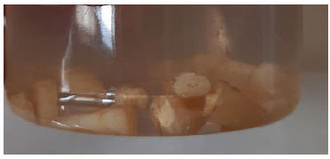
The sealing ability was done on 120 extracted human single-rooted teeth in 6 groups based on a pilot study: 20 sealer samples prepared from each of the groups (A, B, C, D, E) and 20 commercial AH Plus sealer samples as a control group. The procedure was performed in such a way that the teeth were immersed in 5% sodium hypochlorite for 30 minutes to remove surface debris. The crown of the teeth was cut in such a way that the length of the remaining root in each of the samples was equal to 16 mm. Then, the canals were prepared and instrumented with the Protaper file (Dentsply Maillefer, Switzerland) of the F3 and F2 systems. During cleaning, the canal was washed with 5% sodium hypochlorite. After preparation, the canal was washed with 17% EDTA for 3 minutes to remove the smear layer and the canal was washed again with 5 ml of 5% sodium hypochlorite. Finally, the channel was washed with 5 ml of distilled water and dried with sterile paper towels. The canals were thenfilled with a sealer and a single cone [14]. The materials used in the groups were filled in the canal of the teeth. The teeth were placed in a wet environment (100% humidity) for 24 hours. All tooth surfaces were covered with 2 layers of nail varnish, and the orifices were filled with cyanoacrylate paste (Razi Cement Company, Tehran, Iran). This was done to inhibit microleakage from the channels [21]. The temporary repair material was removed from the access cavity of the samples before preparing the leakage evaluation device. To prepare the leakage evaluation device, a hole was inserted in the plastic stopper of a 10 ml glass vial, and the teeth were inserted through this hole and filled with cyanoacrylate paste in between the plastic stopper. A plastic cylinder was connected to the crown of the plastic stop. The 9.5 ml of double-distilled water was added to a glass vial and 1 ml of 22% Bovine Serum Albumin (BSA) (Sigma Chemical Co, St Laurs, MO, USA) was used for filling the cylinder. All experimental groups were placed in the device at 37 °C for 60 days. During the test period, the water in the glass vial was changed daily, and the BSA tank was refilled (Fig. 1).
The presence of protein was evaluated by a reagent (Coomassive Brilliant Blue) on the 60th day. The change in the color of the protein reagent indicated the existence of leakage. The protein was measured by UV spectro- photometer (Genesys 10, Madison, USA). The test was based on observing the absorption maximum for an acidic solution of Coomassie Brilliant Blue (G-250 Bio-Rad Corporation, Life Science, Ca, USA), which occurs in the range of 465 to 595 nm when bound to the protein. The amount of mass of the BSA that has leaked into the space adjacent to the filler material was evaluated via the absorption rate and calibration curve coefficient [21].
2.3. Statistical Analysis
To make a statistical comparison between the studied groups, a One-way ANOVA test was used, and a significance level was considered at p< 0.05.
2.4. Ethical Considerations
The ethics committee at Tabriz University of Medical Sciences provided the ethical code, and all procedures were carried out after getting the ethical code (IR.TBZMED.VCR.REC.1401.167). We confirm that the Helsinki Declaration has been followed in the study. The written informed consent has been taken from the patients to use their teeth.
| - | n | Mean | SD (±) |
|---|---|---|---|
| AH plus | 10 | 0.126 | 0.040 |
| A | 10 | 0.599 | 0.177 |
| B | 10 | 0.413 | 0.120 |
| C | 10 | 0.134 | 0.058 |
| D | 10 | 0.813 | 0.115 |
| E | 10 | 0.854 | 0.093 |
| Control (-) | 10 | 0 | 0 |
| Control (+) | 10 | 0.672 | 0.134 |
3. RESULTS
All groups, except group C, were significantly different from AH Plus and had less sealing properties (p<0.05). Group C had the best sealing properties compared to other groups, and its sealing effect was similar to AH Plus as a commercial sealer (p>0.05). After group C, group B had the best sealing ability compared with other groups, while the E and D groups had the least sealing ability (Table 2).
4. DISCUSSION
Ina successful root canal treatment, the main goal is to remove microorganisms from the root canal and fill the space inside the canal to prevent possible apical pathosis caused by colonization bacteria [22-24]. Conventional root canal treatment is unsuccessful in some clinical cases. Therefore, root canal surgery is a mandatory procedure. Root-end resection and root-end filling are common surgical procedures where conventional endodontic treatment fails. The ideal root-end filling material has good adhesion to the dentin walls, bioactive promotion of healing and tolerance of the surrounding radicular tissue [25].
The goal of this study was to prepare a new endodontic sealer with different percentages of polycaprolactone (P767 and P787) resin, Caprolactone Methacryloxy Ethyl Ester (CMEE), bioactive glass, zinc oxide, barium sulfate, calcium hydroxide, and calcium phosphate and compare the sealability of them. We chose AH Plus sealer for comparison due to its application in clinical efforts. Since this material has good fluidity, suitable layer thickness and good viscosity, it can be used as a control group in studies related to the properties of new sealers. It is used due to better apical seal, reduced solubility, microretention to root canal dentin, and less retraction [26, 27].
Shahi et al. examined the micro apical leakage of zinc oxide and eugenol sealers, tubli seal and AH. 110 single-rooted maxillary central incisor teeth that were freshly extracted were used. After cutting the crown from the Cemento Enamel Junction (CEJ), the preparation of the canals was done by the step-back method so that file No. 35 was used as the Main Apical File (MAF) and the canals were widened up to file No. 60. They divided the tested teeth into 5 groups (three main groups and two control groups). In each group, gutta-percav, one of zinc oxydoxanol, Tubli Seal, and AH sealers were used to fill the canal, except for the positive control group, where no sealer was used. They applied a dye penetration technique to evaluate the amount of microleakage. The linear measurement of color penetration was done with the help of a stereomicroscope, and the data was studied via the LSD test. The findings exposed that there was no significant difference between zinc oxide and eugenol sealer in the amount of color penetration (P=0.63). However, there was a difference in the amount of color penetration between the AH group and the other two groups, as well as between the positive control group and the test group was significant (P < 0.01). The authors concluded that Sealer Zing Oxidaugenol is not suitable for helping to perform successful root canal treatment [28].
In our study, group C had the best sealing properties compared to other groups and its sealing effect was similar to AH Plus as a commercial sealer (p<0.05). The composition of group C was P767 (25%), P787 (10%), Bioactive glass (21.5%), ZnO (21.5%), and BaSO4 (22%), which showed the best sealing result compared with other groups. The suitable sealing ability of group C is due to the simultaneous presence of two polycaprolactones (P767 and P787) in its composition.
Alani et al. evaluated the sealing capability of a composite of polycaprolactone–phosphate glass base for usage as a root canal obturation material. It displayed good potential as a root-filling material capable of generating a seal in an aqueous surrounding without a sealer [29]. Indeed, they used diverse structures of polycaprolactone–iron phosphate glass in different percentages to apply to root canal ex vivo. They produced standardized root canals in extracted human teeth. Then, they studied the ion release, the teeth for root filling adaptation and precipitate formation (using an electron microscopic device) and the sealing capability of the used materials. In their experiment, this group used teeth filled with GP and ordinary zinc oxide/eugenol sealer. The test results showed that, in some cases, there was sediment formation. In this experiment, all different ionic species were released inversely proportional to the concentration of iron oxide. Also, according to the obtained data, after 7 days of immersion in saline, the tested samples showed significantly (P < 0.001) less sediment compared to the control group.
Lin and coworkers evaluated the sealing ability of a root canal filling material with polycaprolactone-base. The examiners used 66 single-rooted extracted teeth (apical size 45) and then obturated with Resilon. Then, they divided the roots into 3 groups (group 1; without treatment, Groups 2 and 3: apical size 60 using K-files and ProFile, respectively) in randomized version. Then, 4 samples from each group were chosen to test via scanning electron microscopy examination. The observed results showed that the remaining roots from groups 2 and 3 were refilled with Resilon. Then, the microleakage test was conductedusing 16 roots from each group which two roots were controls. They analyzed data statistically by Kruskall-Wallis test. There were no significant differences between the investigational groups (P > 0.05) [30].
Tay et al. filled the apical seal in roots with a polycaprolactone-based filling material in vitro, which showed methacrylate-based sealer was not greater than gutta-percha and a conventional epoxy-resin sealer [31]. They tested the ultrastructural feature of the apical seal, which was conducted usingResilon/Epiphany and gutta-percha/AH Plus. They prepared the single-rooted human extracted teeth via a crown-down method, debrided with NaOCl and EDTA, and obturated with either Resilon/ Epiphany or gutta-percha/AH Plus. They try to test the gaps along canal walls and apical leakage using an electronic microscope via SEM and TEM, respectively. The results for SEM exposed both gap-free regions and gap-containing regions in canals filled with both materials. The data for TEM discovered the existence of silver deposits along the sealer-hybrid layer interface in Resilon/ Epiphany, and between the sealer and gutta-percha in the control groups. The authors stated that a complete hermetic apical seal could not be attained with either root-filling materials.
In our study, it seems that the simultaneous presence of two polycaprolactones, as well as bioactive glasses, ZnO and BaSO4, caused a good sealing effect.
CONCLUSION
Sealers play an essential role in sealing teeth. So, gutta-percha cannot performthis task alone. Sealers can fill the root canals of the teeth well and fix the serious damage caused to them. Note that the doctor's skill in using sealers is very important otherwise your teeth may suffer more damage. Then, the goal of this examination was to formulate and evaluate the sealing ability of a new endodontic sealer with conventional AH Plus sealer. In our study, various compositions of sealer material were prepared, and group C had the best result. According to the consequences of other studies and the outcome of our study, it is better to conduct more studies in this field in order to obtain the best sealer composition that has optimal sealing ability. Further study is focused on evaluating the cell cytotoxicity of the new endodontics sealer such that it has a similar sealing ability to AH Plus with lower cytotoxicity.
AUTHORS’ CONTRIBUTION
All the authors were involved in study conception, data collection, data acquisition and analysis, data interpretation, manuscript writing and manuscript revising. All authors have read and agreed to the published version of the manuscript.
LIST OF ABBREVIATIONS
| BSA | = Bovine Serum Albumin |
| SBS | = Shear Bond Strength |
| CMEE | = Caprolactone Methacryloxy Ethyl Ester |
| CEJ | = Cemento Enamel Junction |
ETHICS APPROVAL AND CONSENT TO PARTICIPATE
The ethics committee at Tabriz University of Medical Sciences provided the ethical code, and all procedures were carried out after getting the ethical code (IR.TBZMED.VCR.REC.1401.167).
HUMAN AND ANIMAL RIGHTS
No animals were used in this research. All procedures performed in studies involving human participants were in accordance with the ethical standards of institutional and/or research committees and with the 1975 Declaration of Helsinki, as revised in 2013.
CONSENT FOR PUBLICATION
The written informed consent has been taken from the patients to use their teeth.
AVAILABILITY OF DATA AND MATERIALS
The raw/processed data needed to reproduce these outcomes can be shared at this time. Also, after publication, the data can be requested from the corresponding author via email.
FUNDING
The thesis was supported by the Vice Chancellor for Research at Tabriz University of Medical Sciences, Tabriz, Iran, under the grant number 68866.

