All published articles of this journal are available on ScienceDirect.

High Concentration Whitening Gel Without Remineralizers: The Importance of Polishing and Fluoridation After Tooth Bleaching
Authors Info & Affiliations
Abstract
Background
This study aimed to evaluate the effects of dental polishing and topical application of neutral fluoride after bleaching with 35% hydrogen peroxide (HP35%) without remineralizing bioactive components in its composition on the surface roughness (SR) of tooth enamel.
Material and Methods
Fifty healthy bovine incisors were divided into five groups (n=10 each): G1, without treatment (only stored in artificial saliva); G2, dental bleaching with HP35%; G3, dental bleaching with HP35% + tooth polishing with diamond paste; G4, tooth whitening with HP35% + topical application of fluoride; G5, dental bleaching with HP35% + tooth polishing with diamond paste + topical application of fluoride. SR readings were taken at times T0 (before treatment) and T1 (after treatment). Data were analyzed using analysis of variance for samples related to Tukey's post hoc test.
Results
The lowest mean roughness was observed in G3 at T1 (0.123 Ra) and the highest mean in G1 at T0 (0.198 Ra). However, the values of all groups at T1 were not statistically different (p˃0.05) from the T0 values.
Conclusion
Coronal polishing and topical application of neutral sodium fluoride did not interfere with the roughness of the enamel bleached with a gel without remineralizing agents in its composition.
1. INTRODUCTION
Tooth bleaching is one of the most common esthetic dental procedures and is considered safe and effective when performed as indicated by the manufacturer [1, 2]. Kothari et al. (2019) stated that the color of the dental substrate is a composition of the enamel, dentin, and pulp, and the lighter tone after tooth whitening is mainly due to the optical properties of the enamel, which starts to reflect light without selective absorption of any wavelengths [3].
There are several methods and products available to whiten teeth, with the active ingredients being hydrogen peroxide (HP) or its stabilized form, carbamide peroxide [4]. HP has a low molecular weight, allowing it to diffuse into the tooth structure and trigger a series of oxidative reactions [2]. This agent decomposes into water and reactive oxygen species, which react with the unsaturated organic molecules responsible for color changes in dental tissues [5, 6].
Several studies have shown that these reactions resulting from an in-office bleaching process can lead to alterations in enamel surface morphology [roughness], microhardness, and chemical properties [5, 7, 8]. Enamel changes occur due to several factors, including bleaching agent composition [9], peroxide concentration [10], pH [11], application time [12], and bleaching protocols used [13]. These surface alterations can lead to greater adherence to biofilms [12] and pigments from beverages [14], making them susceptible to extrinsic discoloration and compromising the effectiveness of the treatment [15].
To obtain a smoother enamel surface, some bleaching gel manufacturers recommend polishing after bleaching [16], which can be performed with abrasive discs and/or polishing pastes [17]. Commercially available polishing pastes are typically composed of a lubricant base, thickener, emulsifier, and micronized diamonds of different particle sizes [17, 18]. Another recommended clinical step is the topical application of neutral fluoride on the enamel after whitening to minimize the transient erosive effect, as the whitening gel acts in a non-specific manner [2].
This study aimed to evaluate the surface roughness of tooth enamel bleached with a high concentration gel (HP35%) without remineralizing bioactives in its composition, followed by polishing with diamond paste and/or neutral fluoride applied.
2. MATERIALS AND METHODS
2.1. Sample Preparation
The teeth used in the research were donated by a cattle slaughter company for meat consumption, so the teeth were extracted after the animals were slaughtered. This project was appreciated and was approved by the Ethics Committee on Animal Use of the Federal University of Para (CEUA/UFPA) under number 4273240320.
The sample size was calculated based on the results of a pilot study. GPower software was used to analyze the data and estimate a sample of 10 specimens per experimental group.
In this study, 50 sound bovine incisor teeth were immersed for one week in a 0.1% thymol solution (A Formula Belém, PA, Brazil) for disinfection, followed by removal of adhered periodontal tissue and prophylaxis with pumice stone paste. The teeth were examined under a stereoscopic magnifying glass (40×) to evaluate the enamel of the middle coronal portion and as an inclusion criterion, the buccal enamel should be free of cracks or structural defects on the enamel surface.
The dental crowns were cut into two transverse sections. The first cut was made at a distance of 15 mm (measured with a digital caliper, DIN 862; Mitutoyo, São Paulo, Brazil) from the cementoenamel junction, parallel to the incisal edge. The second cut was made at a distance of 5 mm from the cementoenamel junction with a metallographic cutter (Isomet 1000, Buehler Ltda, Lake Bulf, Illinois, USA), yielding samples of the middle portion of the dental crown with a height of 10 mm (Fig. 1) [1].
All dental enamel fragments had their buccal surface lightly pressed on a wax sheet (No. 07) to position a polyvinyl chloride ring with a height of 10 mm, which was then filled with a chemically activated acrylic resin (JET, Clássico, Campo), 24 h later, the specimens’ buccal surface was flattened in a semiautomatic polisher (Automet 250, Buehler Ltda, Lake Buff, Illinois, USA) with constant cooling, low speed (200 rpm), and a force of 20 N. The sandpapers used were as follows: #400 for 30 s, #600 for 30 s, #1500 for 1 min, and #2500 for 1 min, with each change of previously used sandpaper.
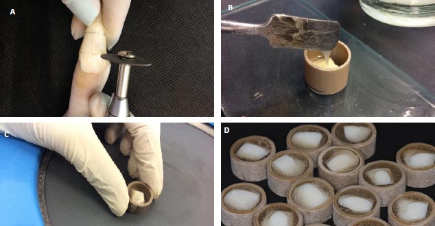
The specimens were then washed in an ultrasonic bath with distilled water to remove the dirt resulting from polishing for 20 min, making them ready for the initial readings. After the initial readings, the specimens were enumerated and randomized into five groups (n=10 each): G1, no treatment (only stored in artificial saliva); G2, tooth bleaching with HP35%; G3, tooth bleaching with HP35% + dental polishing with diamond paste; G4, tooth bleaching with HP35% + topical application of fluoride; and G5, tooth bleaching HP35% + dental polishing with diamond paste + topical application of fluoride.
2.2. Tooth Bleaching
The specimens were bleached with HP35% (Whiteness HP, FGM) according to the manufacturer's instructions. The specimens were stored in artificial saliva (0.7 mmol/L CaCl2, 0.2 mmol/L MgCl2, 4.0 mmol/L KCl, and 20.0 mmol/L HEPES; pH: 7.0) at 37°C throughout the trial period and were renewed daily. A 1-mm thick layer of the bleaching gel (measured by a millimeter probe) was applied to standardize the amount of bleaching gel on the enamel surfaces of the specimens in the test groups. The bleaching gel was shaken every 5 min until the final bleaching time (three applications of 15 min, totaling 45 min per bleaching session). This procedure was repeated three times, with an interval of seven days between each session. After removing the bleaching gel, the teeth were washed, dried, and stored in artificial saliva until the next procedure.
The groups that additionally underwent polishing with diamond paste (Diamond Polish Mint, Ultradent) were polished with a soft felt disk (Diamond Flex, FGM) under constant pressure for 10 s. The specimens were then washed with distilled water using a triple syringe with air and water.
For the groups that additionally underwent topical application of colorless neutral sodium fluoride (DFL), the fluoride gel was placed on the vestibular enamel surface of the specimens with a micro brush for 4 min, and the thickness of the gel was standardized to 1 mm using a millimeter probe. After 4 min, the excess gel was removed using a suction cannula and gently cleaned with gauze. All the specimens were stored in artificial saliva in a biological oven maintained at 37°C.
2.3. Roughness
SR was evaluated using a rugosimeter (SJ-301; Mitutoyo, Los Angeles, CA, United States), and the readings were recorded at two-time points (T0, before the start of treatment and T1, 24 h after the third bleaching session). The parameter adopted to calculate SR was arithmetic roughness, which was determined by the average (µm) of three readings, with a trace limit of 5 mm and a sampling length or cut-off of 0.25 mm.
3. RESULTS
The roughness results measured before and after the intervention showed no statistically significant differences (p˃0.05). The lowest mean was observed in G3 at T1 (0.123 Ra) and the highest mean in G1 at T0 (0.198 Ra) (Table 1).
| Groups | Before Treatment [T0] | After Treatment [T1] |
|---|---|---|
| G1- Negative control | 0.198 [±0.05]a |
0.182 [±0.05]a |
| G2- Tooth bleaching | 0.146 [±0.015]a |
0.188 [±0.056]a |
| G3- Bleaching + polishing | 0.136 [±0.022]a |
0.123 [±0.062]a |
| G4- Bleaching + fluoride | 0.154 [±0.020]a |
0.136 [±0.088]a |
| G5- Bleaching + polishing + fluoride | 0.128 [±0.014]a |
0.144 [±0.048]a |
4. DISCUSSION
In the present study, tooth bleaching and dental polishing using diamond paste and a felt disc alone and/or combined with topical application of 2% neutral sodium fluoride did not influence the topography of dental enamel. The literature on the subject is scarce, and to date, no randomized clinical trials have been conducted with this subject as the main objective [15]. Therefore, laboratory (baseline) studies are required to predict the relevance of clinical treatments. In this in vitro study, the surface topography of the enamel bleached with HP35% without remineralizing bioactive agents and the post-treatments to this therapy (polishing of the bleached enamel and/or topical application of fluoride) were evaluated using a roughness test.
The roughness of intraoral hard tissues and restorative materials can influence plaque retention and pigmentation of dental elements, and a polishing paste seems to be more favorable after bleaching with 35%HP [1, 19]. However, a study by Carvalho et al. states that polishing does not minimize the deleterious effect of bleaching on enamel, but using a polishing paste with a granulometry of 0.5 μm after in-office bleaching may reduce staining over time due to restoration of enamel smoothness [15]. However, the present study did not demonstrate that in-office bleaching alters enamel roughness.
To minimize the adverse effects of peroxide at high concentrations on enamel, manufacturers advocate the application of remineralizing agents such as neutral fluoride topically or incorporated into new formulations [1], including compounds based on calcium or bioactive glass, where they have demonstrated restoration of morphological defects caused by bleaching and reduction of surface irregularities and roughness [4, 15, 20]. In the present study, the bleaching gel used contains no fluorides or other bioactive compounds, with the aim of verifying only the action of peroxide after post-treatment. Even in G2 (bleached only) and G3 (bleached with the addition of topical application of neutral and colorless fluoride), no change in roughness was observed.
Although our study did not demonstrate a difference in treatments, there are still divergences in the literature on the subject, stating that none of the agents have been able to completely reverse the negative effect of the high concentration of HP on the enamel [l4,15,20]. Some manufacturers still recommend polishing after bleaching to obtain a smoother enamel surface [16, 21]. However, in another study, polishing did not prevent staining (which is one of the objectives of polishing) after bleaching, but it restored the smoothness of the base enamel [15].
Regarding the topical application of neutral fluoride, no change was observed in the surface topography of the enamel, and previous studies have shown the union of its ionized form with hydroxyapatite (fluorapatite), forming a flatter surface that is less susceptible to demineralization [4]. It is worth mentioning that the storage medium used in the present study was artificial saliva, which minimized the adverse effects of bleaching on enamel.
Several studies have identified microhardness assessment as one of the parameters used to measure the magnitude of the effects of bleaching procedures [6, 7, 12]. In this connection, we can point out this fact as a limitation of our study. However, this parameter was not evaluated because the hypothesis tested was whether post-treatments affect the enamel surface. We believe that further studies should be carried out because there are a number of new bleaching products in the market that have remineralizing agents [2] and other alternatives that seek to minimize the adverse effects on enamel [17].
CONCLUSION
It is concluded that tooth bleaching with HP35% without remineralizing components in its formulation, as well as post-treatment (enamel polishing and topical application of colorless neutral fluoride), did not influence the roughness of the enamel. Therefore, the recommendation of additional clinical steps after tooth bleaching with high-concentration gels seems to be dispensed with.

