All published articles of this journal are available on ScienceDirect.

FESEM Analysis of Dentin Cleanliness and Erosion at the Apical Third Level after Treatment with Five Irrigating Solutions: An in vitro Study
Abstract
Introduction
For root canal therapy to be successful, thorough debridement and shaping of the root canal system are essential prerequisites. It is understood that removing the smear layer before obturation (filling the canal) can significantly enhance treatment quality. This investigation aimed to assess the efficacy of five distinct irrigating solutions in clearing the smear layer, specifically from the apical one-third of the root canal.
Methods
This in-vitro laboratory study, which utilized Field Emission Scanning Electron Microscopy (FESEM), examined five different solutions: 37% orthophosphoric acid, 12% nitric acid, 42% citric acid, 17% EDTA, and MTAD, a specialized irrigant containing citric acid, an antibiotic, and a surfactant. These solutions were applied to the prepared canals of ninety-six extracted teeth (15 experimental groups and one control group) for 1, 3, or 5 minutes. Following the irrigation period, the tooth specimens were prepared for FESEM analysis. The collected data were then subjected to statistical evaluation using the Mann-Whitney test.
Results
A statistically notable difference, in terms of both smear layer removal and dentinal erosion, was observed in the specimens treated with 37% orthophosphoric acid, 12% nitric acid, and MTAD. In contrast, the other solutions tested did not produce results that were significantly different from the control group.
Discussion
The findings from our research indicate that certain irrigation solutions have a statistically significant effect on smear layer removal when compared to the controls, though this impact was only apparent after a five-minute application period. It is important to highlight, however, that under the experimental conditions of this study, neither 17% EDTA nor 42% citric acid led to any meaningful improvement in smear layer elimination.
Conclusion
Theseresultsunderscore the complex behavior and inconsistent effectiveness of these particular agents, highlight the need for further research to determine the optimal conditions for their application.
1. INTRODUCTION
Many authors have shown that during the shaping of the root canals, a smear layer is created, made of inorganic dentin residues and biological residues derived from the pulp-dentinal complex. This layer of debris may vary in thickness and composition depending on the instrument used in the shaping phase [1]. There has been much discussion about whether to remove this layer from the canal walls [2-6]. Maintaining it intact has been suggested as a means to reduce dentinal permeability and prevent bacterial penetration into the dentinal tubules [2, 3]. Other authors showed that the smear layer contains bacteria and material derived from them, and that therefore its composition prevents the penetration of substances into the intratubular space [4-6]. Finally, it has been suggested that the smear layer should be removed.
1.1. Rationale
We support this hypothesis, as we believe that obtaining an accurate removal of bacteria and their toxins is the clinical basis for avoiding long-term failures. To date, numerous solutions have been tested whose ultimate goal is the removal of the smear layer, but none of them have been effective when compared to the gold standard represented by the use of 5% sodium hypochlorite and 17% EDTA [7]. Apart from irrigating solutions, many NiTi instrument characteristics can modify the production and distribution of debris. These characteristics include the length of the working surface, the diameter of the cross-section, the taper, and the cutting angle of the mechanical tool under consideration [8]. A thicker, more homogeneous smear layer obtained with an instrument with a passive cutting angle will obviously require a more aggressive, longer-lasting action of the irrigating solutions than if a more aggressive instrument were used on the root dentin [9, 10]. Moreover, it has been demonstrated that the depth of irrigation and the type of needle used are two variables that modify the effectiveness of irrigation along the endodontic system [11], especially at the level of the apical third, where, in addition to the anatomical complexity, mechanical instrumentation leaves the endodontic walls covered with a smear layer [12, 13]. Considering all these parameters, there are many studies that have analyzed the removal of the smear layer with different solutions [6, 7], but to date, there are none that compare substances that are even very different from each other. In fact, 37% orthophosphoric acid and 12% nitric acid, often used only for conservative adhesive techniques, as well as MTAD, which represents one of the avant-garde in terms of irrigation, have been compared with solutions such as sodium hypochlorite, EDTA and citric acid, substances that have always been used for endodontic irrigation. All acids were tested at the apical third level, where isthmuses, lateral canals, and cul-de-sacs are difficult to reach even with irrigating solutions [14].
2. METHODS
The methodology for this experiment was adapted from a previously established protocol by Mancini and colleagues [15]. The study received ethical clearance from the Research Ethics Committee at the University of Rome “Tor Vergata” Health Sciences Centre (registration no. 25–30–29) and was conducted in accordance with the Helsinki Declaration. For this investigation, ninety-six human single-rooted mandibular premolars were chosen at random. These teeth were sourced following orthodontic extractions from a cohort of patients from both genders, between 15 and 25 years of age, from whom informed consent had been obtained. The selection criteria required that all teeth be free from any decay, fractures, previous root canal procedures, or fillings. Only teeth with fully formed, undamaged root tips and a total root length of over 15 mm were included. To further refine the selection, radiographs were taken from two angles (buccolingual and mesiodistal) to rule out any teeth exhibiting root curvatures exceeding 5 degrees or signs of canal calcification. Following extraction, the teeth were kept in a 2% thymol solution at ambient temperature and utilized within a week. A laboratory microscope at ×20 magnification (Stemi DV4 Spot; Carl Zeiss, Oberkochen, Germany) was used to scrupulously confirm that all samples met the established inclusion and exclusion standards. After creating an initial access cavity, a size 10 K-file (Dentsply Maillefer, Ballaigues, Switzerland) was carefully advanced down the canal until the tip became just perceptible at the apical foramen. To ensure uniformity, all teeth were shortened to a standard length of 15 mm by making a perpendicular cut to their long axis with a water-cooled, high-speed diamond disc. To mimic a clinical environment, the root apices were sealed with a flowable composite material; a size 10 K-file was temporarily placed in the canal to prevent composite intrusion. To maintain consistency across all samples, a Pro-Train system (Simit Dental, Mantova, Italy) was employed for all tooth preparation steps. The tooth samples were randomly assigned the control group (with 6 teeth) or one of fifteen experimental groups (with 6 teeth each). All canals were shaped using ProTaper Ni-Ti rotary files (Dentsply Maillefer) according to the manufacturer's guidelines, continuing until the F4 file reached the established working length (WL). To maintain cutting efficiency, each individual file was used on a maximum of four teeth. Between each instrument change, the canals were flushed with 3 mL of a 5.25% sodium hypochlorite (NaOCl) solution heated to 37°C (Chematek SpA, Rome, Italy). The patency of the apical foramen was re-established with a size 10 K-file after every shaping step. Following the shaping sequence, each canal received a 1-minute rinse with 3 mL of 17% EDTA (Chematek SpA), followed by flushing with another 3 mL of 5.25% NaOCl at 37°C. The final stage involved the application of the distinct irrigation protocols for each experimental group, as specified in Table 1. The irrigants were delivered into the canals using a 30-gauge syringe needle (NaviTip; Ultradent, South Jordan, UT), which was positioned 1 mm shy of the full working length. To conclude the preparation, all samples were given a final flush with 5 mL of distilled water and were subsequently dried using sterile paper points.
| Group | Time and Irrigating Solution |
|---|---|
| Group A | 1’ Ortophosphoric acid 37% |
| Group B | 3’ Ortophosphoric acid 37% |
| Group C | 5’ Ortophosphoric acid 37% |
| Group D | 1’ Nitric acid 12% |
| Group E | 3’ Nitric acid 12% |
| Group F | 5’ Nitric acid 12% |
| Group G | 1’ Citric acid 42% |
| Group H | 3’ Citric acid 42% |
| Group I | 5’ Citric acid 42% |
| Group L | 1’ EDTA 17% |
| Group M | 3’ EDTA 17% |
| Group N | 5’ EDTA 17% |
| Group O | 1’ MTAD |
| Group P | 3’ MTAD |
| Group Q | 5’ MTAD |
| Group CTR | No final treatment |
2.1. Preparation for Scanning Electron Microscopy
The removal of the endodontic smear layer from instrumented root canals was evaluated using field emission scanning electron microscopy (FESEM). The process involved several key steps [7, 16]:
• Grooving the roots: Each root was longitudinally grooved on its external surface using a diamond disc. This was done carefully to facilitate fracture without damaging the root canals.
• Splitting the roots: The grooved roots were split in half with a chisel. To maintain the integrity of the endodontic canal walls and prevent obscuring fragments, a ProTaper F4 gutta-percha cone was inserted into the root canal before splitting.
• Selecting and coding specimens: For each root, the half that provided the most visible and unobstructed view of the endodontic wall was selected. This chosen half was then coded.
• Mounting: Finally, the coded specimens were securely mounted on metal stubs, ready for FESEM analysis.
To conclude the preparation sequence, the specimens were dried and then examined with a field-emission scanning electron microscope (SUPRA 35; Carl Zeiss SMT, Oberkochen, Germany). The microscope was operated with a gun voltage of 5 kV and a working distance of approximately 11 mm. These specific parameters were selected to prevent excessive electrical charging of the samples. For imaging, a secondary electron detector (SE2) was utilized, as the primary focus of the analysis was the surface topography of the canal walls. For every tooth, five micrographs were captured at standardized locations within the canal (at the very tip, and at 1, 3, 5, and 8 mm from the apex). These images were taken at three distinct magnifications: 300x, 500x, and 1000x.
2.2. Smear Layer Removal Assessment
The evaluation was conducted independently by two observers who were blinded to the experimental groups. Before beginning the official assessment, the observers were calibrated by jointly examining 20 initial specimens. The kappa test was used to assess intra-examiner (consistency of a single observer) and inter-examiner (consistency between two observers) reliability for the microscopic evaluation [17].
The two parameters considered in this study were the degree of intra- and peritubular patency and the degree of root wall erosion resulting from the action of the acids used during pre-treatment. To standardize the research, a 1000x magnification was chosen. To describe the degree of patency and erosion, we used the values described in Tables 2 and 3, codified by Torabinejad et al. [18]. The results were attributed using a double-blind method. The patency and erosion values obtained from the analysis of samples observed under FESEM were then statistically analyzed using the Mann-Whitney test.
| Value | Smear Layer Removal | Visual |
|---|---|---|
| 0 | Complete | Absence of smear layer on canal walls, dentinal tubules free |
| 1 | Partial | Absence of smear layer on canal walls, dentinal tubules full of debris |
| 2 | None | Presence of smear layer on canal walls, dentinal tubules covered |
| Value | Erosion | Visu |
|---|---|---|
| 0 | None | Normal tubules |
| 1 | Middle | Peri-tubular dentin eroded |
| 2 | Severe | Inter-tubular dentin eroded |
3. RESULTS
3.1. Statistical Analysis and Results Summary
Following the methodology of da Costa Lima et al. [16], we used conventional descriptive statistics to summarize quantitative data and non-parametric tests-specifically, Kruskal-Wallis analysis of variance and the Mann-Whitney U test-to evaluate group differences. Multiple comparisons were performed using Bonferroni, Scheffe, and Sidak post-hoc tests. A p-value of 0.05 was set as the threshold for statistical significance. All statistical analysis was performed using STATA software (v12.1).
3.2. Patency and Smear Layer Removal
We compared 15 treatment groups with a control group based on the degree of patency, with significance values ranging from p < 0.05 to p < 0.0001 (Table 4). Several groups showed no significant difference (p > 0.05) from the control group in smear-layer removal, including all groups treated with 42% citric acid (G, H, I), 1-minute nitric acid (D), and all groups treated with 17% EDTA except the 5-minute EDTA group (N). Key findings from the comparisons include:
| F | vs. | CTR | p <0,05 |
| D | vs. | A | p <0,05 |
| D | vs. | E | p <0,05 |
| F | vs. | C | p <0,05 |
| N | vs. | H | p <0,05 |
| N | vs. | L | p <0,05 |
| N | vs. | M | p <0,05 |
| Q | vs. | O | p <0,05 |
| A | vs. | CTR | p <0,005 |
| E | vs. | CTR | p <0,005 |
| D | vs. | B | p <0,005 |
| N | vs. | G | p <0,005 |
| N | vs. | CTR | p <0,005 |
| B | vs. | CTR | p <0,0005 |
| P | vs. | CTR | p <0,0005 |
| C | vs. | CTR | p <0,0001 |
| Q | vs. | CTR | p <0,0001 |
The 1-minute orthophosphoric acid group differed significantly from both the 1-minute nitric acid group (p < 0.05) and the control group (p < 0.005).
The 3-minute nitric acid group differed from the 5-minute nitric acid group (p < 0.05).
The 3-minute orthophosphoric acid group differed significantly from the 1-minute nitric acid group (p < 0.005) and the control group (p < 0.0005).
The 5-minute orthophosphoric acid and 5-minute MTAD groups showed the highest statistical difference compared with the control group (p < 0.0001).
The 5-minute EDTA group (N) demonstrated superior smear-layer removal compared to the 1-minute citric acid group (G, p < 0.005), and also outperformed groups H (3-minute citric acid), L (1-minute EDTA), and M (3-minute EDTA) (p < 0.05).
3.3. Erosion
Erosion values showed significance ranging from p < 0.02 to p < 0.0001. Groups that did not differ significantly from others included all groups treated with 42% citric acid (G, H, I), 1-minute nitric acid (D), and 17% EDTA (L, M, N) (Table 5).
The 1-minute orthophosphoric acid group and the control group differed significantly from the 3-minute nitric acid group (p < 0.02), and from the 3-minute orthophosphoric acid and 5-minute nitric acid groups (p < 0.001).
| E | vs. | A | p <0,02 |
| E | vs. | CTR | p <0,02 |
| C | vs. | F | p <0,005 |
| B | vs. | A | p <0,001 |
| B | vs. | CTR | p <0,001 |
| F | vs. | A | p <0,001 |
| F | vs. | CTR | p <0,001 |
| Q | vs. | O | p <0,001 |
| Q | vs. | CTR | p <0,0005 |
| C | vs. | CTR | p <0,0001 |
The control group also differed significantly from the 5-minute orthophosphoric acid group (p < 0.0001).
The 5-minute inorganic acid groups (C and F) differed from each other at p < 0.005.
The 5-minute MTAD group was significantly more erosive than both the 1-minute MTAD group (p < 0.001) and the control group (p < 0.0005).
4. DISCUSSION
As a result of the present study, an ideal irrigating solution that meets all the desired requirements is not currently available. Numerous solutions are in use, each with limitations: some are highly effective at dissolving the organic matrix but show minimal effect on the inorganic component [19]. Among the most commonly used irrigating solutions, sodium hypochlorite meets the required criteria better than others, although there remains some debate regarding its optimal concentration and temperature of use.
The inorganic acid-based solutions examined in this study did not produce fully satisfactory results. Both orthophosphoric acid and nitric acid provided good cleaning in the middle third of the root, consistent with previous literature [17, 19, 20], but they were less effective in the apical third, where cleaning is most critical. At the middle and coronal thirds, our samples showed results comparable to those reported in the literature, indicating that our sample preparation technique is suitable for this type of study (Fig. 1). Near the apex, instrumentation residues partially or completely occluded the tubules, along with concretions of varying shapes and sizes-likely associated with saline precipitates-and bacterial microorganisms of different types (Fig. 1) [21].
Considering that this is an in vitro study conducted under ideal conditions, one can only speculate about clinical outcomes, where working conditions are often more challenging. The apex of the tooth serves as both an entry and exit point for potential contamination and houses most lateral and accessory canals, with considerable anatomical variability. Effective cleaning and sealing of this region are essential for successful root canal therapy, as incomplete treatment may lead to frustrating relapses.
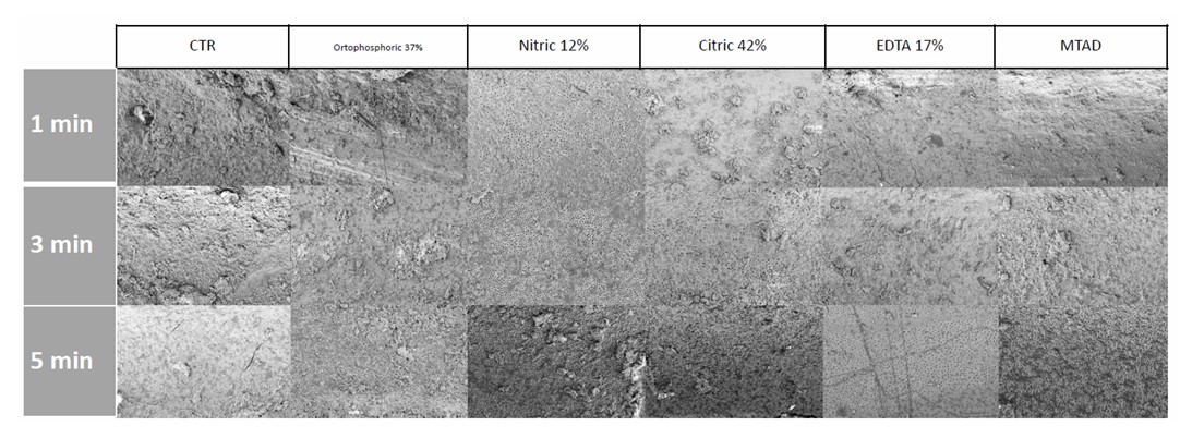
FESEM representative images.
According to our in vitro results, orthophosphoric acid leaves the root surface with residual debris at different application times. Statistical analysis revealed no significant differences in patency between the 1-minute treatment and the control, while 3- and 5-minute treatments achieved significant improvements. Erosion also differed significantly from the control for the 3- and 5-minute treatments (p < 0.001 and p < 0.0001, respectively).
For nitric acid, its effect on the smear layer appears to selectively remove the inorganic component. This preference is confirmed in the rare cases where inter-tubular dentin was completely eroded, usually due to incomplete removal of the overlying smear layer. However, nitric acid demonstrated significant activity only when applied for more than 1 minute, as shorter durations did not achieve statistically significant improvements in patency.
5. STUDY LIMITATION
The main limitation of the present study is the relatively small number of specimens and the use of a single shaping file size (.40), especially considering that minimally invasive endodontics (MIE) can now be performed using a variety of shaping files and advanced metallurgical designs.
CONCLUSION
This study highlights a striking observation: only a minimal number of root canals meet all the ideal requirements. However, this does not appear to be the primary cause of endodontic treatment failure. These findings suggest the need to re-evaluate the factors that determine long-term, predictable success, and to consider whether treatment efficacy is more closely related to reducing bacterial load than to the traditionally emphasized aspects of instrumentation, irrigation, pre-treatment, and three-dimensional obturation.
AUTHORS’ CONTRIBUTIONS
The authors confirm contribution to the paper as follows: M.M., L.C.: Study conception and design, Data collection, Analysis and interpretation of results, Draft manuscript. All authors reviewed the results and approved the final version of the manuscript.
LIST OF ABBREVIATIONS
| EDTA | = Ethylenediaminetetraacetic |
| MIE | = Minimally Invasive Endodontics |
ETHICS APPROVAL AND CONSENT TO PARTICIPATE
The study received ethical clearance from the Research Ethics Committee at the University of Rome “Tor Vergata” Health Sciences Centre, Italy (registration no. 25–30–29).
HUMAN AND ANIMAL RIGHTS
All procedures performed in studies involving human participants were in accordance with the ethical standards of institutional and/or research committee and with the 1975 Declaration of Helsinki, as revised in 2013.
AVAILABILITY OF DATA AND MATERIALS
All the data and supporting information are provided within the article.
ACKNOWLEDGEMENTS
Declared none.

