All published articles of this journal are available on ScienceDirect.

Prevalence and Patterns of Periodontal Diseases Among Patients Attending Dental Clinics in Al-Nasiriya City, Iraq: A Cross-Sectional Study
 iD
iD
Authors Info & Affiliations
Abstract
Background
Periodontal diseases have become a serious public health problem all over the world. The objective of this study was to evaluate the extent and severity of periodontal diseases among patients in Al-Nasiriya city and its association with some selected demographic and clinical variables, using the 2017 World Workshop classification of periodontal diseases.
Methods
A cross-sectional survey study was carried out on 2,243 adult (20-75 years) patients of both sexes from different central and peripheral districts of Al-Nasiriya city. The periodontal condition was assessed with the Community Periodontal Index for Treatment Needs (CPITN) and conventional periodontal parameters, such as probing pocket depth (PPD), bleeding on probing (BOP), and clinical attachment level of loss (CAL). Measurements of the periodontium were performed in six sites around all teeth examined.
Results
The prevalence of periodontal diseases (gingival bleeding, calculus, and periodontal pockets) in the study cohort was very high. Gingivitis was identified in 330 (14.9%) patients, while periodontitis was present in 1,913 (85.1%). Clinically healthy periodontal tissues were present in only 194 cases (8.8%). Both prevalence and severity of periodontitis increased with age and were substantially associated with smoking.
Discussion
The current reorganized analysis confirms a substantial burden of periodontal disease in Al-Nasiriya city, characterized by age-related progression and significant associations with modifiable systemic and behavioral risk factors. These findings underscore the urgent need for integrated preventive strategies tailored to the Iraqi population.
Conclusion
Periodontitis prevalence in adults of Al-Nasiriya city was high. Age and being a smoker are two host factors of great importance that affect periodontal health. These findings emphasize the necessity of further preventive strategies and public health programs for awareness and risk factor control of periodontal disease in this segment of society.
1. INTRODUCTION
Periodontal diseases are persistent inflammatory disorders of the gingiva and surrounding supporting tissues of the teeth, such as the periodontal ligament and alveolar bone. These diseases represent a continuum from the reversible plaque-induced gingivitis to periodontitis, which involves irreversible loss of attachment, resorption of alveolar bone, and ultimate loss of teeth [1, 2]. Periodontal diseases are a global health problem due to their widespread prevalence and the chronic destruction of tissues [3, 4].
Epidemiological data show that periodontal diseases are highly prevalent among adult populations worldwide and demonstrate exponentially increasing prevalence and severity with age [5, 6]. One of the most common non-communicable diseases worldwide, according to the Global Burden of Disease Study, was severe periodontitis, and it was also a leading cause of years lived with disability due to oral conditions [7]. Existing studies illustrate the high level of human, social, and economic cost resulting from periodontal diseases placed on individuals and health-care systems [8, 9].
The epidemiology and distribution of periodontal diseases differ greatly between populations and geographic areas. Several population-based studies in Europe, Asia, and North America have reported substantial variations in disease distribution, extent, and severity [10-12]. Differences in age distribution, socioeconomic status of the population, oral hygiene practices, and access to preventive and curative dental care have been linked to such variation [13, 14].
Precise estimation of the prevalence of periodontal disease has been for a long time rendered difficult due to variation in diagnostic criteria and methods used during examination. Previous epidemiologic studies often were based on partial-mouth examination methods or non-uniform case definitions, which might have resulted in an underestimation of disease burden and difficulty comparing among studies [15, 16]. By introducing the 2017 World Workshop classification, a huge progress was achieved by providing standardized case definitions based on stages and grades of diseases and adding severity, complexity, and risk of progression [17]. This model has enhanced reliability and comparability of modern epidemiologic data [8].
Several confirmed risk indicators have been reported to affect prevalence and severity of periodontal disease. One of the most important modifiable risk factors is tobacco smoking, and it greatly alters the progression of the disease and level of tissue destruction [18]. Systemic disorders like diabetes mellitus, obesity, osteoporosis, and hormonal changes also predispose to an increased degree of susceptibility and severity of periodontal disease [19-21]. Furthermore, sex- and age-related disparities in the profile of periodontal disease have been well documented, consistently indicating both biological and behavioral parameters impacting oral health [22-24].
In addition to oral morbidity, the involvement of periodontal diseases in systemic health conditions, via common inflammatory pathways, is well established. There is evidence that periodontitis and systemic diseases, especially diabetes mellitus, interact in a bidirectional manner. Thus, the concept of periodontal health in relation to overall health and healthy ageing is being increasingly focused upon [25-27].
Although periodontal disease has been thoroughly studied around the world, there is little epidemiological data about its prevalence, especially in some Middle East countries. Recent Iraqi studies that have applied the 2017 classification system showed a high prevalence of periodontitis, stressing that more standardized epidemiological surveys are required in this area [28, 29].
Developing local data is critical for recognizing unique disease patterns in populations to be able to implement successful strategies of prevention and treatment. Thus, the current study aimed to establish the prevalence, distribution, and severity of periodontal diseases in a sample of patients attending dental clinics in Al-Nasiriya city, Iraq, using the 2017 World Workshop classification system, and their distribution in relation to age groups and gender.
2. MATERIALS AND METHODS
2.1. Study Design and Setting
A cross-sectional study was planned to assess the prevalence and patterns of periodontal health and disease in patients attending dental clinics of Al-Nasiriya City, Iraq. Data were collected between October 2021 and December 2023 at the Department of periodontology Al-Nasiriya Dental Specialized Center. The study protocol was designed based on well-defined guidelines for epidemiological as well as periodontal research standards [10, 28-30].
2.2. Study Population and Sample Selection
The study population comprised 2,243 participants aged 20–75 years, including 1,237 males and 1,006 females. Participants were randomly selected from patients attending the dental center during the study period for reasons unrelated to Temporomandibular Disorders (TMD). Individuals with systemic diseases known to affect periodontal status were excluded to minimize potential confounding [31-33]. Written informed consent was obtained from all eligible participants before the clinical examination.
These include that the sample comprised 2,243 individuals, which is deemed sufficient, as a small sample size would reduce statistical power to assess important differences and associations between periodontal disease outcomes and major characteristics such as age and smoking. Since the large sample size provides estimates of prevalence with low sampling error, given the prevalence reported here, this data set is valuable. In addition, it enables credible subgroup analyses by demographic and clinical characteristics. The sample size helps ensure robust generalizable finds and improves analysis of the strength of observed associations within the target population, supporting the study’s cross-sectional objectives. The study was reported in accordance with the Sex and Gender Equity in Research (SAGER) guidelines.
2.3. Clinical Periodontal Examination
Full-mouth periodontal examinations were carried out by calibrated examiners. Bleeding on probing (BOP), probing pocket depth (PPD), and clinical attachment loss (CAL) were recorded at six sites per tooth using standard periodontal probes (a University of North Carolina periodontal probe) [34]. Diagnosis of periodontal disease and case definitions were based on the 2017 World Workshop Classification of Periodontal and Peri-Implant Diseases and Conditions, co-developed by the American Academy of Periodontology (AAP) and the European Federation of Periodontology (EFP) [17]. Presence of periodontal health in intact, reduced, and stable periodontium was defined as BOP <10% with all probing depths ≤3 mm without attachment loss related to previous or current periodontitis [35].
2.4. Periodontal Indices and Case Definitions
Screening for periodontal treatment need was performed according to the recommendations of the World Health Organization using the Community Periodontal Index of Treatment Need CPITN [36]. Examination was carried out using a WHO-periodontal probe (0.5-mm ball tip) and one black band from 3.5 to 5.5 mm, cleaned teeth marginally between 14 and 16 hours after the last meal. The probe was gently (squash) advanced circumferentially around the sextant, and the maximum score per sextant was noted.
The CPITN index classifies periodontal condition using three key features: bleeding on probing, presence of dental calculus, and periodontal pocket depth [36]. Periodontal Examination (BPE) scores (0–3) were also recorded. Periodontitis was diagnosed when interproximal attachment loss was observed [12].
2.5. Radiographic Assessment Methodology
Although the clinical examination is a valuable but limited indicator for assessing periodontal status, radiography provides additional information regarding alveolar bone level and pattern of loss that cannot otherwise be determined. As part of the periodontal evaluation, standardized digital periodontal and/or panoramic radiographs were reviewed in this study [17]. Radiographs were examined for the presence, extent, and severity of alveolar bone loss (vertical and horizontal bone defects, furcation involvement, other radiographic signs of periodontal destruction).
Bone loss was evaluated by assessing the distance between the cemento-enamel junction (CEJ) and the alveolar crest, measured as negative values, interpreted alongside clinical parameters such as probing pocket depth (PPD) or clinical attachment loss (CAL). Implant periodontitis classification and staging were based on consensus diagnostic criteria using radiographic findings. Radiographs were evaluated by trained examiners, with all readings under standardized viewing conditions; questionable findings were reviewed by a senior clinician to maintain consistency.
2.6. Periodontitis Classification: Stage, Grade, and Extent
Extent of periodontitis was defined as localized (when less than 30% of the teeth were involved) and generalized (30% or more of the teeth involved) [12]. A molar–incisor configuration was apparent if first molars and incisors exhibited most of the bone loss [17].
The severity of periodontitis was classified according to clinical attachment loss (CAL) at the most advanced site: Stage I (CAL <2 mm), Stage II (CAL 3–4 mm), and Stage III–IV (CAL ≥5 mm), with further modifications according to tooth loss caused by periodontitis [17].
The disease grade was used for estimate the progression rate by calculating the ratio between radiographic bone loss and patient age, adjusted for smoking habit and diabetes mellitus [1, 17]. Ratios <0.5 indicated slow progression (Grade A), 0.5–1.0 moderate progression (Grade B), and >1.0 rapid progression (Grade C).
Disease status was also classified into three groups (stable periodontitis, remission of the disease, and unstable periodontitis) based on the presence of bleeding on probing and residual periodontal pocket depths after therapy [12].
2.7. Inclusion Criteria
Participants were included in this study if they fulfilled the following conditions:
- Individuals between 20 and 75 years old.
- Both males and females.
- The inhabitants of Al-Nasiriya city central/Peripherical dwellings.
- Setting: Patients seen at selected dental clinics during the study period.
- Persons who had given informed consent to participate.
- Had at least one natural tooth to enable periodontal evaluation.
2.8. Exclusion Criteria
Study participants were excluded if they met any of the following criteria:
- Patients less than 20 years or more than 75 years.
- Edentulous patients (absolutely toothless), since periodontal evaluation could not be performed.
- Individuals with a periodontal treatment history within the last 3–6 months.
- People with conditions that may alter their ability to undergo periodontal examination (e.g., acute oral infections, severe systemic disease).
- Stating that they declined participation or showed incomplete clinical data.
2.9. Statistical Analysis
Statistical analyses were performed using Python software. Continuous variables, including BOP, PPD, and CAL, were summarized using descriptive statistics (means and standard deviations). Frequencies and percentages were calculated for categorical variables. Correlation analyses were conducted to assess associations among periodontal parameters. Multinomial logistic regression was performed to assess the relationship between periodontal disease stage and potential predictor variables such as age, sex, tooth brushing frequency, smoking status, Diabetes Mellitus (DM), and hypertension [1, 33, 37]. The model was applied in Python using the MNLogit function from the package statsmodels. Categorical variables were treated as binary indicators based on dummy coding.
Model fit was evaluated using log-likelihood values, pseudo R2 statistics, and likelihood ratio tests [38]. The statistical significance of individual predictors was determined using regression coefficients, z-values, and corresponding p-values. A two-sided p-value of less than 0.05 was considered statistically significant.
3. RESULTS
3.1. Baseline Socio-demographic and Clinical Characteristics of the Study Population
Table 1 presents the distribution of participants according to their socio-demographic characteristics and medical determinants. A total of 2,243 subjects were included in the study. Regarding age, nearly equal proportions were observed, with 1,100 participants (49%) aged <40 years and 1,143 (51%) aged >40 years. The majority of the sample were Arabic (2,122; 94%), while non-Arabic individuals constituted a small minority (121; 5.3%).
| Age (Year) | |||
|---|---|---|---|
| 20-40 | 40-60 | 60-75 | |
| Healthy | 100 (4.5%) | 44 (2%) | 50 (2.3%) |
| Gingivitis | 142 (6.4%) | 99 (4.5%) | 89 (4%) |
| Stage 1 | 128 (5.7%) | 90 (4%) | 88 (4%) |
| Stage 2 | 102 (4.5%) | 145 (6.5%) | 168 (7.5%) |
| Stage 3 | 90 (4.1%) | 142 (6.4%) | 170 (7.7%) |
| Stage 4 | 69 (3%) | 120 (5.3%) | 164 (7.3%) |
In terms of educational level, 982 participants (43.9%) were highly educated, 810 (36%) had a normal educational level, and 451 (20%) were not educated. Concerning systemic health conditions, 262 subjects (11.6%) were diabetic, 196 (8.7%) had hypertension, 102 (4.54%) were obese, and 41 (1.82%) reported osteoporosis.
Gender distribution showed a predominance of males (1,237; 55.14%) compared with females (1,006; 44.98%). Regarding smoking habits, 523 participants (23.3%) had never smoked, 304 (13.55%) were former smokers, and 410 (18.29%) were current smokers. Overall, the sample demonstrated a balanced age distribution, moderate-to-high educational attainment, and a noticeable proportion of systemic conditions and smoking exposure.
3.2. Distribution and Age-related Patterns of Periodontal Status Among the Study Population
This cross-sectional study included 2,243 individuals aged 20–75 years and assessed periodontal status according to the 2018 classification system. As illustrated in Table 2, only 194 participants (8.65%) were periodontally healthy, indicating a low prevalence of optimal periodontal health in the study population. Gingivitis was diagnosed in 330 individuals (14.71%). Periodontitis was the most prevalent condition, affecting 1,476 participants (65.80%). Among these, 306 (13.64%) were classified as Stage I, 415 (18.50%) as Stage II, 402 (17.92%) as Stage III, and 353 (15.74%) as Stage IV, demonstrating a considerable burden of moderate to advanced disease.
| Determinants | No. | |
|---|---|---|
| Age | <40 years >40 years |
1100 (49%) 1143 (51%) |
| Ethnicity | Arabic Non-Arabic |
2122 (94%) 121 (5.3%) |
| Education | High educated Normal educated Not educated |
982 (43.9%) 810 (36%) 451 (20%) |
| Medical condition | Diabetics Hypertension Obesity Osteoporosis |
262 (11.6%) 196 (8.7%) 102 (4.54%) 41 (1.82%) |
| Gender | Male Female |
1237 (55.14%) 1006 (44.98%) |
| Smoking | Never smoked Former smoker Smoker |
523 (23.3%) 304 (13.55%) 410 (18.29%) |
Stratified by age, different age-related trends were identified. The largest proportion (51.55%) of the 194 healthy people was in the age range 20–40 years; 22.68% and 25.77% are between ages 41–60 and 61–75 years, respectively. Likewise, the most common age group observed with gingivitis is 20–40 years (43.03%), followed by 41–60 years (30.00%) and 61–75 years (26.97%).
The distribution pattern of stage I peaked in the 20-40 age group (41.83%), followed by the age groups between 41-60 (29.41%) and 61-75 years old (28.76%). Moderate to severe degrees of periodontitis, on the other hand, reported an evident rise with increasing age. Of the cases of stage II periodontitis, 24.58% were recorded in the age between 20 and 40 years, while 34.94% and 40.48% were recorded in the ages between 41–60 and between the ages of 61–75, respectively. A similar distribution pattern was detected for stage III, with 22.39% in the 20–40-year age group, compared with 35.32% and 42.29% in the 41–60- and 61–75-year groups, respectively. The highest concentration of severe disease was noted in Stage IV, with nearly half of the cases (46.46%) occurring in participants aged 61–75 years, compared to 33.99% in those aged 41–60 years and only 19.55% in the youngest age group.
Overall, these findings indicate that periodontal health was more common among younger adults, whereas the prevalence and severity of periodontitis increased progressively with age, with advanced stages predominantly affecting individuals aged 61–75 years. This age-dependent gradient highlights the cumulative and progressive nature of periodontal tissue destruction over time.
3.3. Descriptive Statistics and Prevalence
As presented in Table 3, a total of 565 participants were analyzed for the periodontal parameters BOP, PPD, and CAL. The mean BOP score was 0.52 ± 0.16, with values ranging from 0.05 to 0.99. The median value was 0.54, and the interquartile range (IQR) extended from 0.43 (25th percentile) to 0.64 (75th percentile), indicating that half of the participants had BOP scores within this moderate-to-high bleeding range.
| BOP | PPD | CAL | |
|---|---|---|---|
| Count | 565 | 565 | 565 |
| Mean±SD | 0.52±0.16 | 4.35±0.60 | 4.18±0.97 |
| Minimum | 0.05 | 3.5 | 3 |
| 25% | 0.43 | 4 | 4 |
| 50% | 0.54 | 4 | 4 |
| 75% | 0.64 | 5 | 5 |
| Maximum | 0.99 | 6 | 9 |
For PPD, the mean value was 4.35 ± 0.60 mm, with a minimum of 3.5 mm and a maximum of 6.0 mm. The median was 4 mm, and the IQR ranged from 4 to 5 mm, reflecting generally moderate pocket depths in the sample. CAL showed a mean of 4.18 ± 0.97 mm, with values between 3.0 mm and 9.0 mm. The median CAL was 4 mm, with an IQR of 4–5 mm, suggesting a moderate degree of attachment loss in most participants.
According to Table 4, prevalence analysis showed that 50.97% of participants had BOP scores greater than 0.5, indicating a high prevalence of clinically significant gingival inflammation. The sex distribution was nearly balanced, with 53.81% males and 46.19% females included in the study.
| Prevalence | |
|---|---|
| BOP > 0.5 | 50.97% |
| Male | 53.81% |
| Female | 46.19469% |
3.4. Correlation Analysis
The correlation matrix presented in Table 5 demonstrates positive associations among all periodontal parameters. BOP was moderately correlated with PPD (r = 0.330257) and CAL (r = 0.390322). The correlation between PPD and CAL was weaker (r = 0.174904).
| BOP | PPD | CAL | |
|---|---|---|---|
| BOP | 1 | 0.330257 | 0.390322 |
| PPD | 0.330257 | 1 | 0.174904 |
| CAL | 0.390322 | 0.174904 | 1 |
Correlation analysis presented in Table 5 demonstrated statistically significant positive associations among all periodontal parameters (p < .001 for all comparisons). BOP showed a moderate positive correlation with PPD (r = 0.330257) and with CAL (r = 0.390322), indicating that increased gingival bleeding was associated with deeper periodontal pockets and greater attachment loss. A weaker but significant positive correlation was observed between PPD and CAL (r = 0.174904), suggesting that although pocket depth and attachment loss are related, the strength of their association was lower compared to the relationships involving BOP. Overall, BOP exhibited the strongest association with CAL (Table 6).
| BOP | PPD | CAL | |
|---|---|---|---|
| BOP | 0 | 7.63E-16 | 5.29E-22 |
| PPD | 7.63E-16 | 0 | 2.91E-05 |
| CAL | 5.29E-22 | 2.91E-05 | 0 |
These relationships are visually illustrated in Fig. (1) (correlation heatmap), which highlights stronger color intensity between BOP and CAL compared to PPD and CAL, indicating that bleeding on probing demonstrated the strongest association with attachment loss among the examined variables.
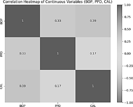
Correlation heatmap displaying the associations between BOP, PPD, and CAL scores.
3.5. Multinomial Logistic Regression
A multinomial logistic regression analysis was conducted to evaluate the association between age, brushing frequency, smoking status, and diabetes status with periodontal disease stages, using Stage 1 as the reference category. The model converged successfully (Log-Likelihood = –1206.4) and was statistically significant (LLR χ2 = 294.2, p < .001). The Pseudo R2 value of 0.1087 indicates that approximately 10.87% of the variance in periodontal disease stages was explained by the included predictors, suggesting a modest but statistically meaningful contribution of these risk factors to disease severity.
3.6. Multinomial Logistic Regression and Correlation Analysis Results
The multinomial logistic regression model identified several significant predictors of periodontal disease stages when Stage 1 was used as the reference category.
3.6.1. Stage 2 vs. Stage 1
As shown in Table 7, increasing age was strongly and positively associated with Stage 2 periodontal disease compared with Stage 1 (B = 1.39, SE = 0.12, t = 11.90, p < .001; 95% CI: 1.16–1.62). This indicates that older individuals had significantly higher log-odds of being classified in Stage 2. The magnitude and direction of this effect are illustrated in Fig. (2).
| Periodontal disease = 0 | Coef. | SE | t | P>|t| | [0.025 | 0.975] |
|---|---|---|---|---|---|---|
| Constant | -2.50 | 0.73 | -3.41 | 0.00 | -3.94 | -1.06 |
| Age | 1.39 | 0.12 | 11.90 | 0.00 | 1.16 | 1.62 |
| Brushing | -0.64 | 0.12 | -5.32 | 0.00 | -0.87 | -0.40 |
| sex_Male | -0.03 | 0.21 | -0.17 | 0.87 | -0.44 | 0.37 |
| smoking_Smoker | 0.35 | 0.17 | 2.02 | 0.04 | 0.01 | 0.69 |
| diabetes_Non-Diabetic | -1.36 | 0.39 | -3.53 | 0.00 | -2.12 | -0.60 |
| hypertension_Non-Hypertensive | 0.85 | 0.57 | 1.49 | 0.14 | -0.27 | 1.97 |
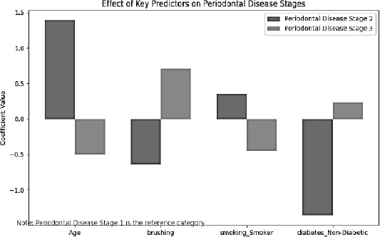
Predictor effects on periodontal disease Stages 2 and 3 relative to Stage 1. Positive coefficients increase the likelihood of advanced stages, while negative coefficients decrease it.
Brushing frequency was negatively associated with Stage 2 (B = −0.64, SE = 0.12, t = −5.32, p < .001; 95% CI: −0.87 to −0.40), indicating that more frequent brushing reduced the likelihood of moderate periodontal disease.
Smoking showed a positive and statistically significant association (B = 0.35, SE = 0.17, t = 2.02, p = .044; 95% CI: 0.01–0.69), suggesting smokers were more likely to be in Stage 2 than non-smokers.
Regarding systemic conditions, Diabetes status was significant; non-diabetic individuals had lower odds of Stage 2 disease (B = −1.36, SE = 0.39, t = −3.53, p < .001; 95% CI: −2.12 to −0.60), implying that diabetic patients were more susceptible to moderate periodontal destruction. Sex and hypertension were not statistically significant predictors (p > .05), as presented in Table 7.
3.6.2. Stage 3 vs. Stage 1
For Stage 3 disease, brushing frequency demonstrated a significant positive association (B = 0.70, SE = 0.13, z = 5.33, p < .001), suggesting higher reported brushing frequency was associated with increased odds of Stage 3. This finding, illustrated in Fig. (2), may reflect reverse causation or residual confounding.
In contrast, Smoking showed a negative association with Stage 3 (B = −0.45, SE = 0.21, z = −2.19, p = .029), indicating smokers were less likely to be in Stage 3 relative to Stage 1.
3.6.3. Correlation Analysis
Pearson correlation analysis demonstrated a weak but statistically significant positive correlation between age and periodontal disease stage (r = .13, p < .05), indicating that disease severity slightly increased with age.
Brushing frequency showed a very weak positive correlation with periodontal disease (r = .047, p = .036), suggesting minimal linear association.
A weak negative correlation was observed between age and brushing frequency (r = −.057, p < .05), indicating that older individuals reported slightly less frequent brushing.
Overall, as supported by Table 7, Fig. 2, and Fig. 3, age emerged as the most consistent predictor of periodontal disease progression, while behavioral variables demonstrated weaker and occasionally contradictory associations.
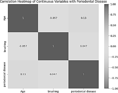
Correlation heatmap displaying the associations between age, brushing, and periodontal disease scores.
4. DISCUSSION
The baseline socio-demographic profile of the study population demonstrated a relatively balanced age distribution, with nearly equal proportions of participants below and above 40 years. The predominance of Arabic ethnicity and the moderate-to-high educational attainment reflect the demographic characteristics of Al-Nasiriya city. A noticeable proportion of participants presented with systemic conditions, including diabetes and hypertension, alongside considerable exposure to smoking. These baseline characteristics are clinically relevant, as systemic diseases and behavioral factors are well-established modifiers of periodontal disease progression [3, 4]. Despite global improvements in oral health awareness over recent decades [39, 40], disparities persist in developing regions, often influenced by socioeconomic factors and access to preventive care.
From a broader epidemiological perspective, the observed distribution of systemic and behavioral determinants aligns with international reports emphasizing the multifactorial nature of periodontal diseases. The Global Burden of Disease Study [7] and international consensus statements [8, 41, 42] highlight that age, metabolic disorders, and tobacco exposure significantly contribute to the worldwide burden of periodontitis. Therefore, understanding these baseline characteristics is fundamental when interpreting disease distribution within this Iraqi cohort.
The distribution of periodontal status revealed that only 8.65% of participants were periodontally healthy, whereas the majority exhibited gingivitis or varying stages of periodontitis. Severe forms (Stages III and IV) affected more than one-third of individuals, with a clear progressive increase across age groups. This age-related escalation mirrors findings from European cohorts using CDC/AAP classifications, although the prevalence in Al-Nasiriya appears comparatively higher [29, 43]. Such differences may be attributed to limited preventive programs, reduced access to specialized periodontal care, and suboptimal oral hygiene behaviors within the population [11, 31].
The strong age-dependent gradient observed in this study confirms the cumulative and progressive nature of periodontal tissue destruction. Advanced stages predominated among older individuals, supporting longitudinal evidence demonstrating that prolonged exposure to plaque biofilm and systemic inflammatory burden leads to greater attachment loss over time [44]. Similar prevalence patterns have been reported in the United States, Europe, and recent Iraqi data employing the updated 2018 classification system [6, 28], reinforcing the public health relevance of these findings.
The descriptive periodontal parameters further illustrate the inflammatory burden within the cohort. Mean values for bleeding on probing, probing pocket depth, and clinical attachment loss indicated moderate-to-advanced periodontal destruction. Notably, over half of participants exhibited BOP scores exceeding 0.5, and males demonstrated slightly higher bleeding prevalence than females. Previous epidemiological studies have reported similar sex-based tendencies, often attributing them to behavioral differences in oral hygiene practices and healthcare utilization [22, 23, 45]. However, such differences may diminish after adjusting for confounding variables.
The moderate central tendency values for PPD and CAL reflect clinically meaningful periodontal breakdown across the sample. These findings correspond with established biological mechanisms whereby persistent gingival inflammation precedes connective tissue degradation and attachment loss [2]. The distribution of these clinical parameters confirms that the disease burden extends beyond mild inflammation to include structural periodontal destruction in a substantial proportion of participants.
Correlation analysis demonstrated statistically significant positive associations among BOP, PPD, and CAL. Bleeding on probing showed the strongest relationship with clinical attachment loss, emphasizing its value as an indicator of active inflammatory burden. These findings are consistent with previous literature demonstrating that gingival bleeding reflects inflammatory activity that may precede measurable tissue destruction [35]. The visual heatmap representation further highlighted stronger associations between inflammatory and destructive parameters.
The biologically plausible relationship among these variables aligns with contemporary models of periodontal pathogenesis [2]. Although the correlation between pocket depth and attachment loss was weaker, it remained statistically significant, suggesting that while related, these parameters represent distinct aspects of periodontal breakdown. Together, these results validate the internal consistency of the clinical measurements and support their use in epidemiological assessment.
The multinomial logistic regression model provided additional insight into predictors of disease severity. Increasing age was the most consistent determinant, significantly elevating the likelihood of more advanced stages. Smoking demonstrated a significant association with Stage 2 disease, confirming its established role as a major modifiable risk factor [6, 18, 46]. Tobacco exposure is known to impair host immune response, alter vascular function, and exacerbate periodontal tissue destruction [47].
Diabetes also emerged as a significant predictor, supporting the well-documented bidirectional relationship between periodontal inflammation and metabolic dysregulation [19, 25, 48]. Although brushing frequency showed variable associations across stages, previous longitudinal research emphasizes that effective plaque control, rather than brushing frequency alone, is critical for maintaining periodontal stability [49]. The modest pseudo R2 value indicates that only part of disease variability is explained by the included predictors, reinforcing the multifactorial etiology of periodontitis involving microbial, host, behavioral, and systemic determinants [38, 50].
Oral hygiene behaviors within this cohort were notably suboptimal, with a high proportion of individuals rarely using dental floss and limited adherence to twice-daily brushing. National survey comparisons suggest minimal improvement in periodontal parameters over time [14]. In an aging society such as Al-Nasiriya, advanced periodontal destruction may compromise masticatory efficiency, nutritional intake, and overall quality of life, particularly among elderly individuals [34]. These findings highlight the need for strengthened preventive education, early detection programs, and improved access to periodontal care.
Several limitations must be considered. The cross-sectional design precludes causal inference; behavioral variables were self-reported, and detailed radiographic data necessary for comprehensive grading assessment were unavailable. Nevertheless, the application of the contemporary 2018 periodontal classification enhances comparability with recent international research [17, 21]. Overall, this reorganized analysis confirms a substantial burden of periodontal disease in Al-Nasiriya city, characterized by age-related progression and significant associations with modifiable systemic and behavioral risk factors. These findings underscore the urgent need for integrated preventive strategies tailored to the Iraqi population.
5. STUDY LIMITATIONS
Some limitations of this study should be acknowledged in the interpretation of the findings. The cross-sectional design limits the ability to infer causal relationships between periodontal disease and related risk factors, including age and smoking. The study was conducted on only those patients attending dental clinics, which could induce selection bias and limit the generalization of results to the wider community. Furthermore, potential confounding factors such as socioeconomic status, oral hygiene habits and systemic diseases were not fully evaluated. This method of using a partial view may be useful in practice but underestimates the severity of disease relative to full mouth diagnostic approaches.
CONCLUSION
In conclusion, this study demonstrates that periodontal diseases were very common among adult patients in Al-Nasiriya city, where the number of individuals with periodontitis was significantly higher than those with healthy periodontal tissues. Both the severity and incidence of disease progress with age and are closely linked to smoking, highlighting the contribution of modifiable behavioral risk factors. These results point to a large public health burden and suggest a lack of awareness and preventive care. Hence, this population warrants the implementation of comprehensive preventive strategies, promotion of oral health education, and early screening and intervention programs to reduce the impact of periodontal diseases in the population.
AUTHOR’S CONTRIBUTIONS
The authors confirm contribution to the paper as follows: study conception and design: Jabbar A. A. Al-Saaidi, Ihsan A. Kumail and Husam S. Ismail; data collection: Husam S. Ismail and Nejood B. Ghetran; analysis and interpretation of results: Jabbar A. A. Al-Saaidi and Ihsan A. Kumail; draft manuscript: Husam S. Ismail and Nejood B. Ghetran. All authors reviewed the results and approved the final version of the manuscript.
LIST OF ABBREVIATIONS
| AAP | = American Academy of Periodontology |
| BOP | = Bleeding on Probing |
| BPE | = Basic Periodontal Examination |
| CAL | = Clinical Attachment Loss |
| CDC | = Centers for Disease Control and Prevention |
| CEJ | = Cementoenamel Junction |
| CI | = Confidence Interval |
| CPITN | = Community Periodontal Index of Treatment Needs |
| DM | = Diabetes Mellitus |
| EFP | = European Federation of Periodontology |
| IQR | = Interquartile Range |
| LLR | = Log-Likelihood Ratio |
| MNLogit | = Multinomial Logit Model |
| PPD | = Probing Pocket Depth |
| R2 | = Coefficient of Determination |
| SD | = Standard Deviation |
| SE | = Standard Error |
| TMD | = Temporomandibular Disorders |
| WHO | = World Health Organization |
ETHICS APPROVAL AND CONSENT TO PARTICIPATE
This research was authorized by the Scientific and Ethical Committee of the College of Dentistry, Al-Ayen Iraqi University, Al-Nasiriya City, Thi-Qar Governorate, Iraq (No. 1183).
HUMAN AND ANIMAL RIGHTS
All procedures involving human participants were performed in compliance with the principles of the Declaration of Helsinki.
CONSENT FOR PUBLICATION
All participants provided written informed consent before enrollment. Confidentiality of the participants was strictly protected, and data were used for research purposes only.
ACKNOWLEDGEMENTS
Declared none.