All published articles of this journal are available on ScienceDirect.

Feldspathic Porcelain Laminate Veneers: A Step-by-step Clinical Case Report

Abstract
Introduction
Dental professionals strive to achieve natural, optical, and biomimetic results in esthetic rehabilitation. The predictability of veneer therapy is based on shape, color, and advanced clinical proficiency.
Case Presentation
This clinical case report describes the anterior esthetic management of a 60-year-old female patient presenting aesthetic concerns related to pre-existing direct composite restorations. A diagnostic wax-up followed by an intraoral mock-up was performed to facilitate three-dimensional evaluation of the proposed restorative outcome and to obtain patient approval prior to definitive treatment. Complete anterior rehabilitation was obtained using lithium disilicate glass-ceramic veneers. Laboratory procedures were conducted in close collaboration with a skilled dental technician to achieve precise surface texture, translucency gradients, and anatomical contouring consistent with adjacent natural teeth. Adhesive luting protocols were meticulously implemented to optimize bonding performance and marginal integrity. Postoperative assessment revealed high patient satisfaction in terms of both aesthetics and function.
Conclusion
The present case underscores the clinical relevance of biomimetic restorative strategies and preoperative visualization tools in enhancing treatment predictability, functional integration, and patient-centered outcomes in contemporary aesthetic dentistry.
1. INTRODUCTION
The increasing demand for anterior aesthetic treatments has driven the evolution of more conservative restorative approaches. Traditionally, full-coverage crowns were considered the most reliable option; however, they require extensive removal of healthy tooth structure, potentially compromising pulpal and periodontal health. The introduction of enamel etching techniques by Buonocore marked a turning point in adhesive dentistry, leading to the development of advanced bonding systems and composite materials [1-5]. Although direct resin composites are effective for minor aesthetic corrections, their long-term stability is limited due to wear, discoloration, and marginal degradation [5, 6]. For this reason, they are often reserved for less demanding or temporary solutions. Porcelain veneers, first introduced in the early 20th century, have been the subject of many encouraging articles on the follow-up of such a therapy [7-9]. Lately, porcelain veneers have undergone significant advancements, particularly with the introduction of reliable etching and bonding protocols [10]. Today, they represent a minimally invasive alternative to crowns, requiring significantly less tooth reduction while providing superior aesthetic outcomes. Contemporary ceramic laminate veneers are recognized for their superior optical properties and long-term aesthetic stability. Their glazed, non-porous surfaces contribute to excellent biocompatibility and reduced plaque accumulation, thereby promoting periodontal health when appropriate oral hygiene is maintained [10, 11]. Compared to full-coverage restorations, veneers represent a more conservative treatment option, typically requiring only 3% to 30% reduction of the tooth structure. The clinical success of these restorations is further supported by advancements in silane coupling agents and adhesive resin cements, which optimize the bond between ceramic and enamel substrates [10, 11]. Their advantages include excellent biocompatibility, resistance to staining, and favorable interaction with periodontal tissues. Modern adhesive systems, combined with silane coupling agents and improved luting materials, have further enhanced their clinical performance [2, 3]. Indications for ceramic veneers encompass a broad spectrum of aesthetic and functional concerns, including dental wear (such as abrasion), coronal fractures, developmental anomalies (e.g., altered tooth morphology or interdental spacing not requiring full crowns), diastemas, and discrepancies in tooth size or shape that cannot be resolved through orthodontic treatment alone. Additionally, veneers are indicated in cases of severe intrinsic discoloration unresponsive to bleaching or micro-abrasion, as well as for minor occlusal corrections, including the management of rotated or protruded teeth. Nevertheless, several limitations must be considered. The treatment protocol is time-intensive and typically requires multiple clinical and laboratory stages. Ceramic veneers are inherently fragile prior to adhesive luting, complicating handling and placement. Repair procedures are often challenging, and provisionalization may present additional clinical difficulties. Furthermore, the procedure is irreversible due to the need for enamel reduction, and it is associated with higher costs compared to direct composite restorations. The technique is also highly sensitive, requiring meticulous clinical execution. Contraindications include cases with insufficient enamel substrate, such as heavily restored or severely worn dentition (due to bruxism and clenching), as well as teeth exhibiting pronounced discoloration that cannot be adequately masked by thin ceramic layers. Non-vital teeth may also present challenges due to potential discoloration and compromised structural integrity. A comprehensive preoperative assessment is essential to ensure optimal treatment planning and to facilitate minimally invasive preparation. Whenever possible, bonding procedures should be performed predominantly on enamel using adhesive resin cements, as this has been shown to enhance the longevity and predictability of ceramic veneer restorations [12-17].
2. CASE REPORT
The present clinical report describes an upper jaw anterior rehabilitation of a 60-year-old female patient extending from canine to canine, performed using a systematic, stepwise protocol with feldspathic porcelain laminate veneers.
2.1. Informed Consent
As an aesthetic procedure, a full discussion of the benefits, risks, and functional objectives is mandatory. Alternative treatments must be presented, along with a detailed timeline of the clinical steps. Written informed consent was obtained from the patient for this study. Ethical approval and the Helsinki Declaration are not applicable as the patient was treated in a private practice where only informed and signed consent is required.
2.2. Shade Selection
Shade selection is challenging due to the veneer's translucency. he definitive optical outcome is influenced by the interaction among the underlying tooth substrate, the selected luting agent, and the ceramic’s thickness and opacity. To enhance shade matching and laboratory communication, digital photography and instrumental color analysis, such as spectrophotometric devices (e.g., VITA Easyshade), were employed. It should be considered that resin-based luting agents may undergo chromatic alterations during polymerization [18]. Additionally, the clinician must consider the color of endodontically treated teeth, especially those sealed with older bioceramics like Mineral Trioxide Aggregate (MTA) [19].
2.3. Diagnostic Wax-up and Mock-up
A prosthetically driven treatment plan was established through the fabrication of a diagnostic wax-up, allowing three-dimensional visualization of the intended outcome. This step also facilitated the creation of silicone indices, which were subsequently used for provisional restorations and as reduction guides during tooth preparation. The proposed morphology was transferred intra-orally through a mock-up, enabling real-time evaluation of aesthetics, phonetics, and function, as well as patient approval prior to irreversible procedures (Figs. 1–4).
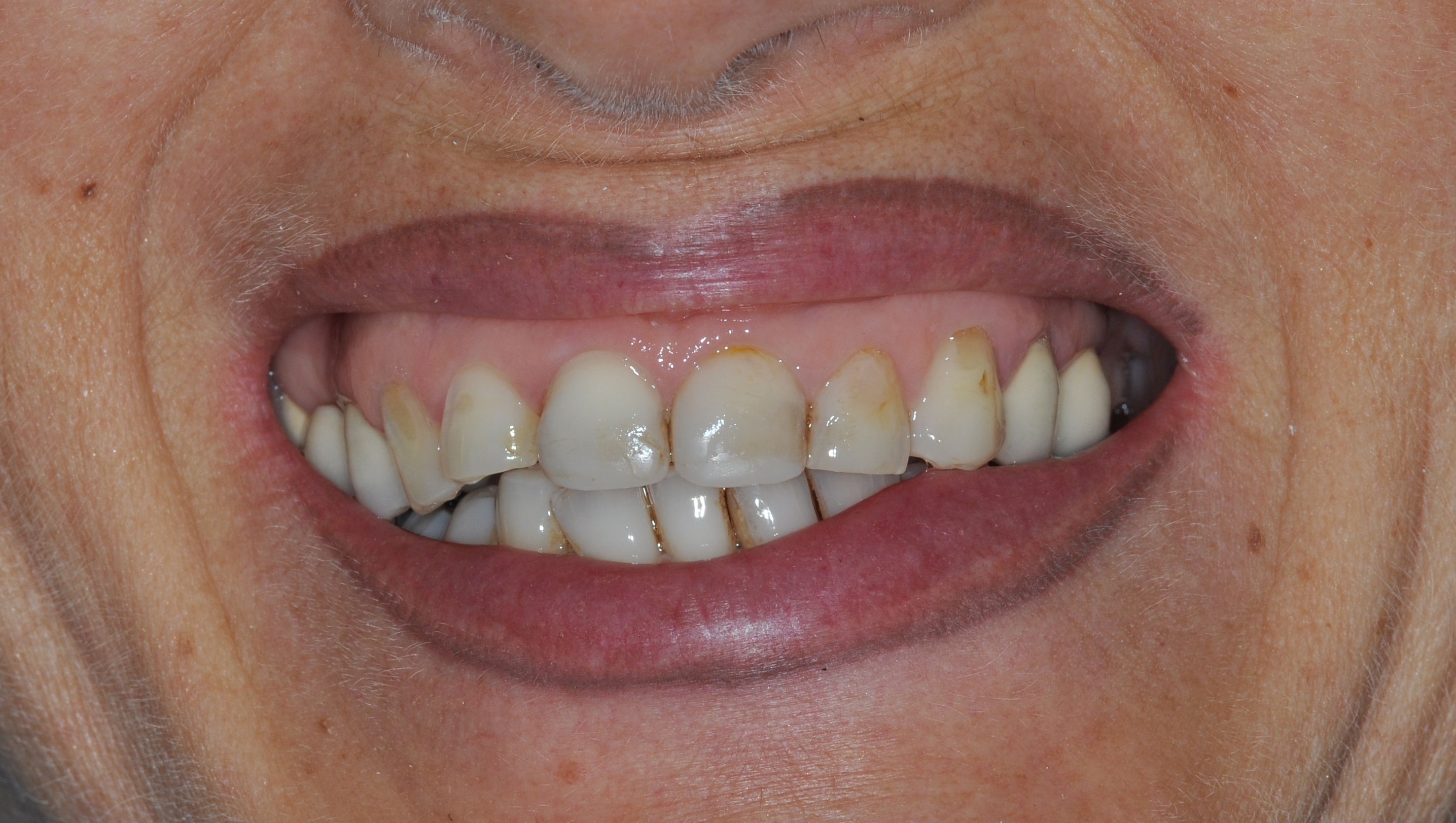
Preoperative.
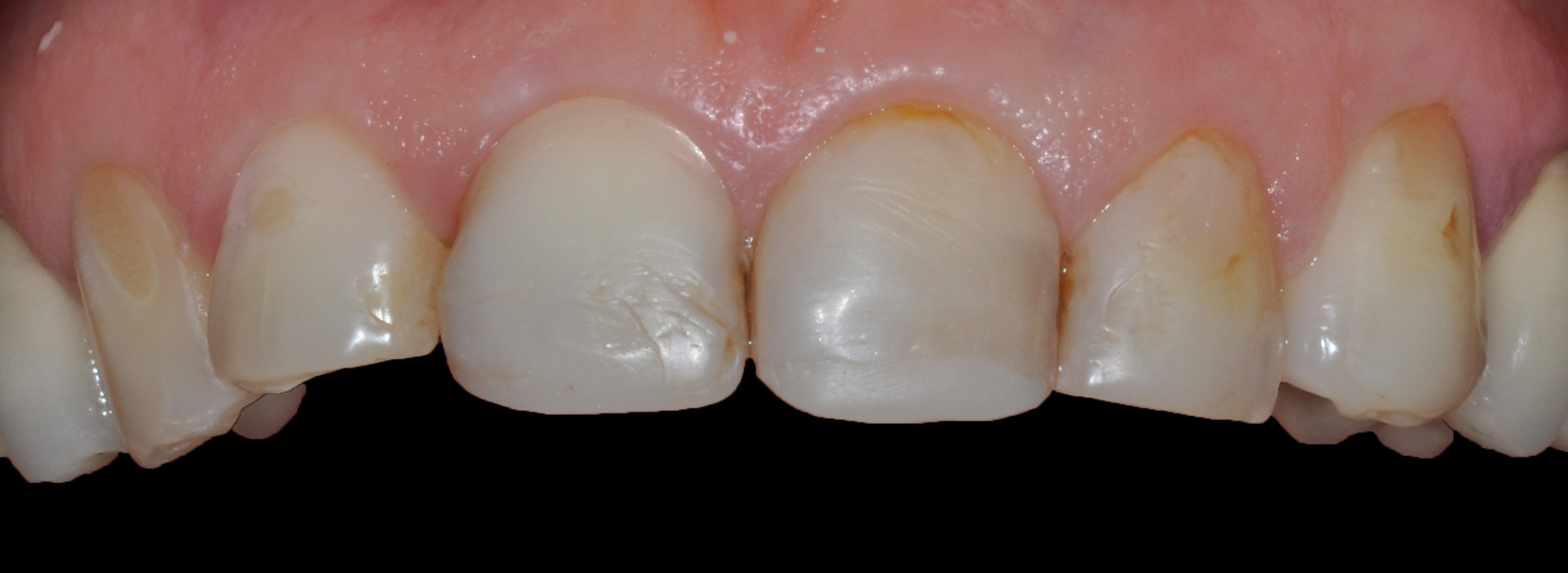
Preoperative. Close-up.
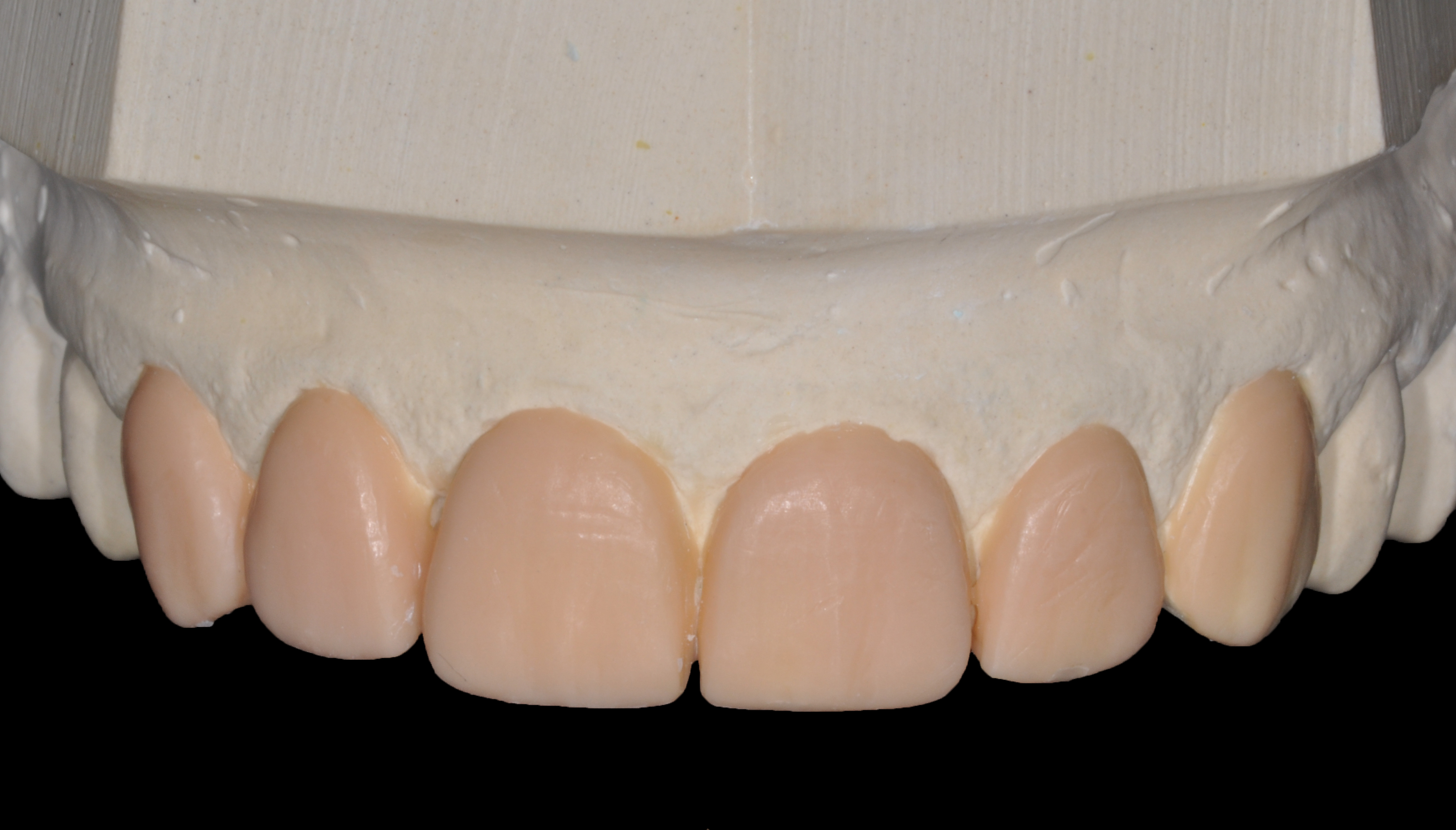
Diagnostic wax-up.
Using a reduced PVS index, a resin temporary from the diagnostic wax-up was created.
2.4. Tooth Preparation
Tooth preparation for ceramic veneers was performed with the objective of preserving enamel while creating optimal conditions for adhesion and material performance. The guiding principles included maintaining preparation within enamel whenever feasible, avoiding sharp internal line angles to reduce stress concentration, establishing clearly defined margins for proper emergence profile, ensuring adequate spatial accommodation for ceramic thickness, and providing precise seating references to facilitate accurate placement.
2.4.1. Vestibular Preparation
Facial reduction of the anterior teeth was executed following a three-plane approach, incisal, middle, and cervical thirds, to respect natural tooth anatomy. Controlled labial reduction was achieved within a range of approximately 0.3 to 0.6 mm, depending on the selected ceramic system (feldspathic porcelain versus lithium disilicate). Three principal techniques were utilized to ensure accurate enamel reduction:
- Freehand preparation, relying on clinical experience and visual assessment.
- Depth-orientation grooves created with calibrated burs to standardize reduction.
- Silicone putty indices derived from the diagnostic wax-up, enabling precise evaluation of reduction volume. Sectioning the index in horizontal planes allowed verification of preparation depth at various vertical levels, ensuring sufficient space for the planned restorative contours (Figs. 5-7).
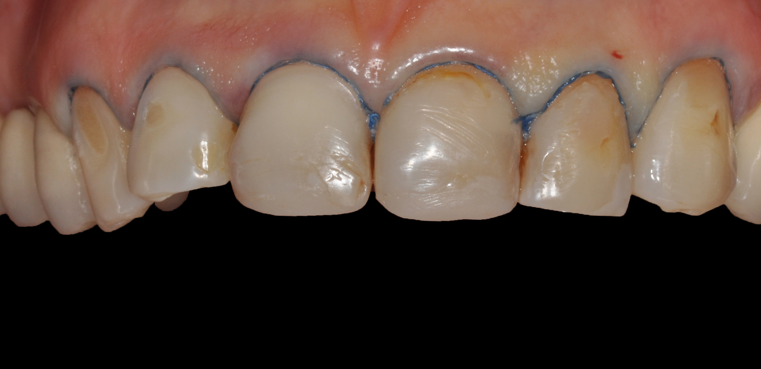
Retraction cord #1 in the gingival sulcus (Ultrapak, Ultradent).
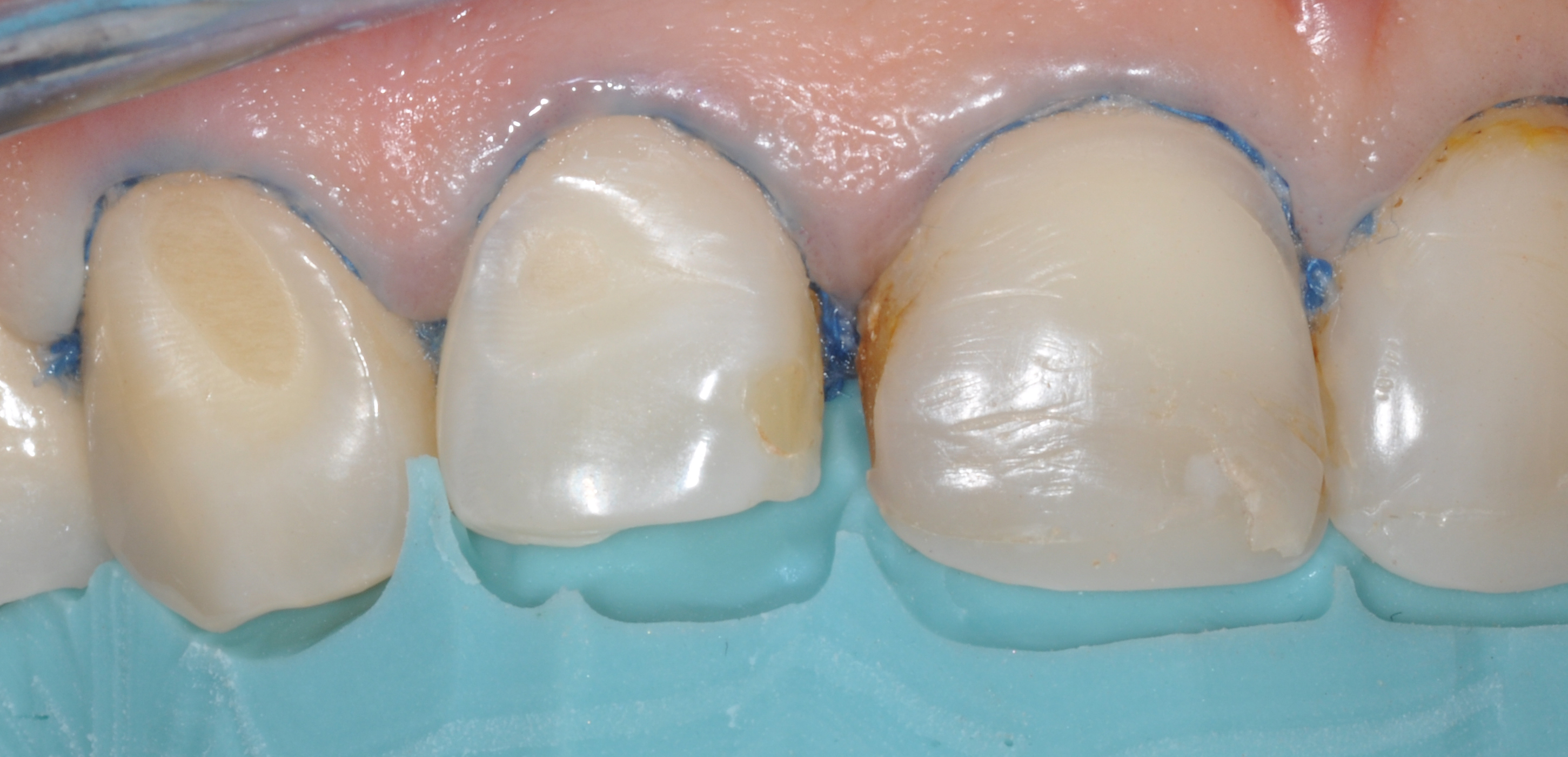
First quadrant PVS reduction index in use.
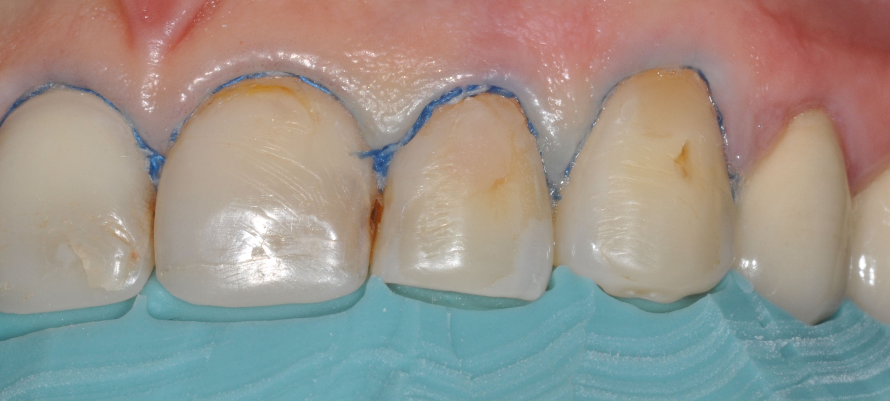
Second quadrant PVS reduction index in use.
2.4.2. Incisal Edge Reduction (Butt-joint Preparation)
Several preparation designs have been described in the literature, ranging from non-incisal (“window” or “feather-edge”) configurations to designs incorporating varying degrees of incisal reduction. In this case, an incisal overlap design was selected, extending from the buccal surface to the palatal aspect, with a reduction of approximately 1.5–2.0 mm. This approach has been advocated in the literature as it enhances the mechanical resistance of the restoration, provides a definitive path of insertion during cementation, and facilitates the laboratory fabrication of natural incisal translucency.
2.4.3. Proximal Preparation
Interproximal extension of the preparation was determined by the desired modification of tooth morphology. When no change in contact relationships was required, the preparation was terminated just facial to the contact area. In contrast, extension through the contact zone was indicated in cases requiring alteration of tooth width or closure of diastemas, allowing for improved control of emergence profile and contact point positioning.
2.4.4. Cervical Margin Preparation
A light chamfer finish line with a depth not exceeding 0.4 mm was established at the cervical region. This margin configuration minimizes the risk of over-contouring, promotes accurate seating of the restoration, and reduces stress concentration within the ceramic. In contrast to full-coverage crowns, subgingival margin placement is generally unnecessary due to the optical blending capability of thin ceramic veneers, often described as a “contact lens effect.” Supragingival margin placement offers multiple advantages, including preservation of enamel for bonding, reduced risk of periodontal irritation, and simplified impression procedures (Figs. 8, 9). Nevertheless, different preparation techniques may be considered (equigingival or minimally subgingival margins).
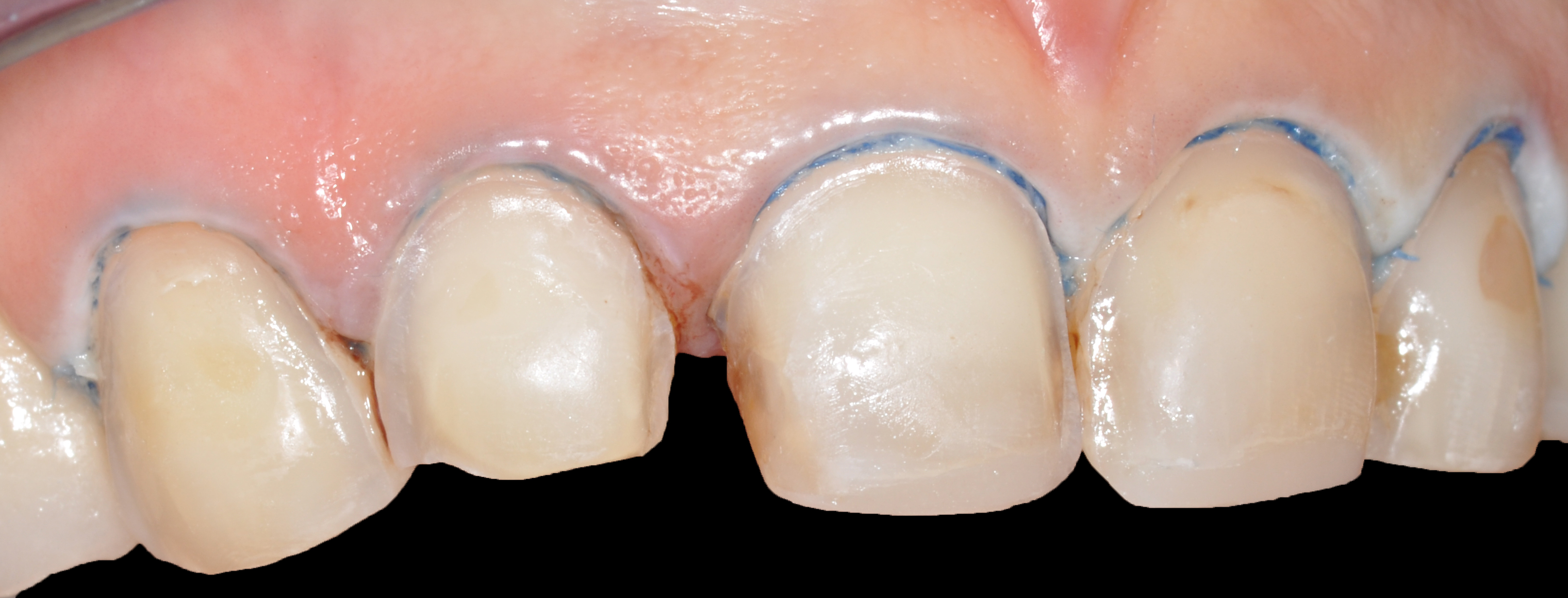
First quadrant teeth preparation (infiltrated Class III restoration will be treated after the cementation of the veneer). Immediate dentine sealing was performed.
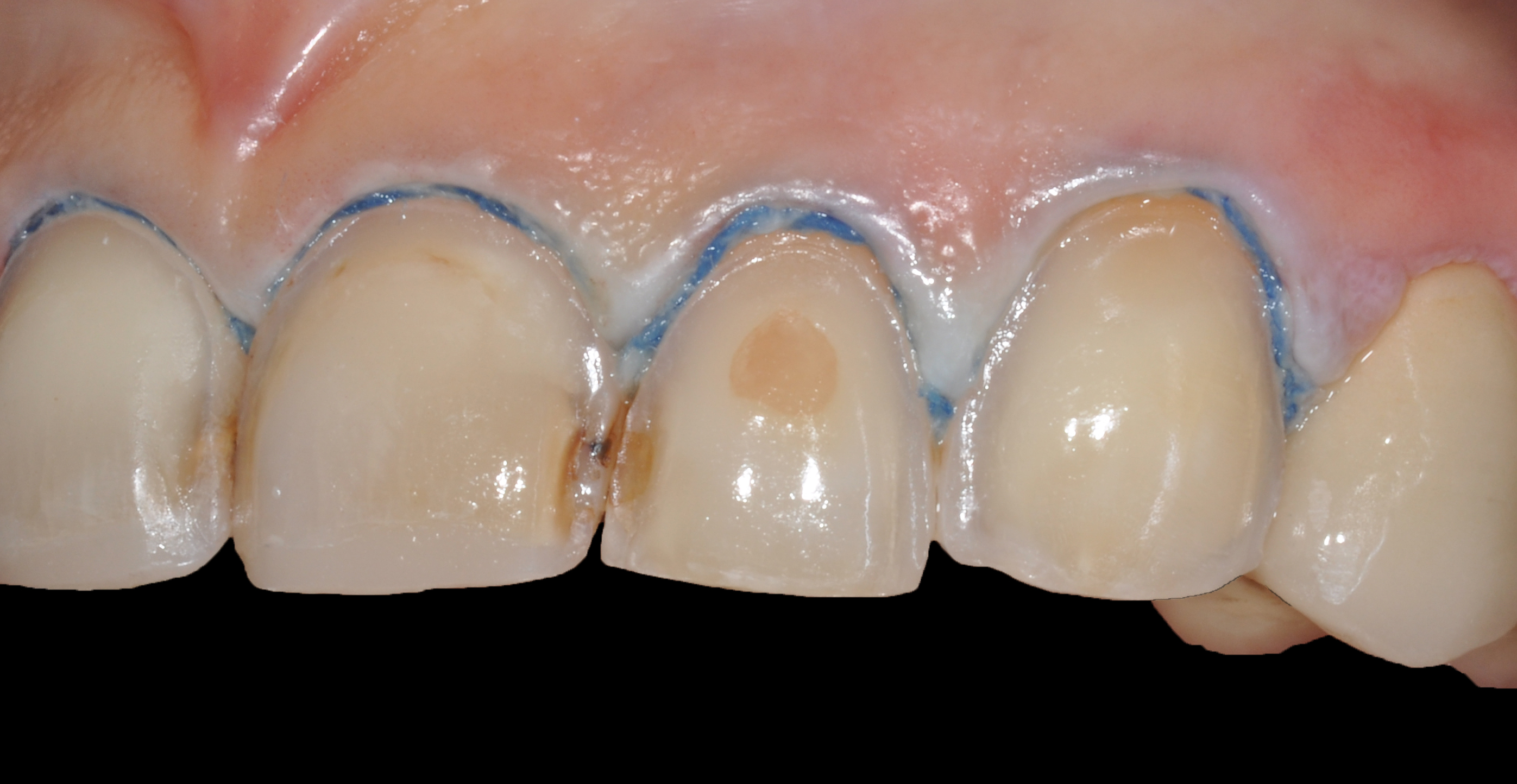
Second quadrant teeth preparation (infiltrated Class III restoration will be treated after the cementation of the veneer). Immediate dentine sealing was performed.
2.5. Facebow Transfer and Bite Registration
Accurate orientation of the incisal plane is critical for laboratory communication. A practical method to achieve this is the use of a “stick bite” or “symmetry bite,” which helps relate the dental midline to the interpupillary line. This can be accomplished by embedding two rigid markers (e.g., sticks) into the bite registration material, providing a visual guide for proper alignment during mounting.
2.6. Temporization
Although provisional restorations are sometimes omitted due to conservative tooth preparation, many clinicians regard this phase as essential. Provisional veneers fabricated from the diagnostic wax-up offer patients the opportunity to preview both function and aesthetics prior to finalization (Fig. 10), facilitating communication and potential modifications.
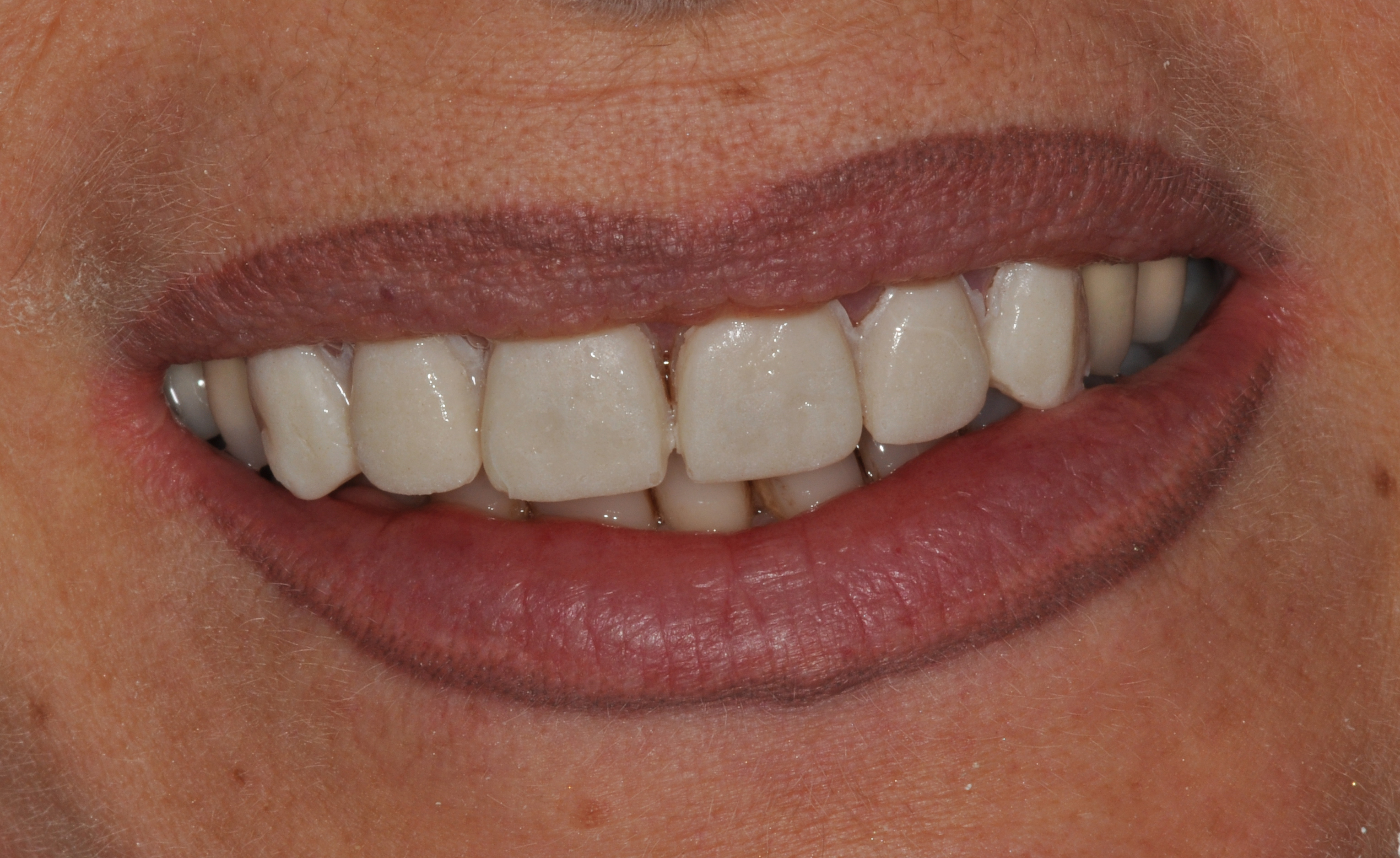
Temp-Bond Clear® (Kerr Dental) was used to cement temporarily.
2.7. Veneer Try-in
Following fabrication (Figs. 11–13), the prepared teeth must be thoroughly cleaned to eliminate any remnants of provisional cement or resin. Each veneer is then evaluated individually to verify marginal fit and adaptation. This assessment may be performed with or without try-in pastes or water; in some cases, avoiding these media can enhance visualization of the margins. Improper seating is often associated with residual debris or excessive proximal contact tightness. After confirming fit and obtaining patient approval of the aesthetic outcome, the restorations can proceed to the bonding stage (Figs. 14, 15).
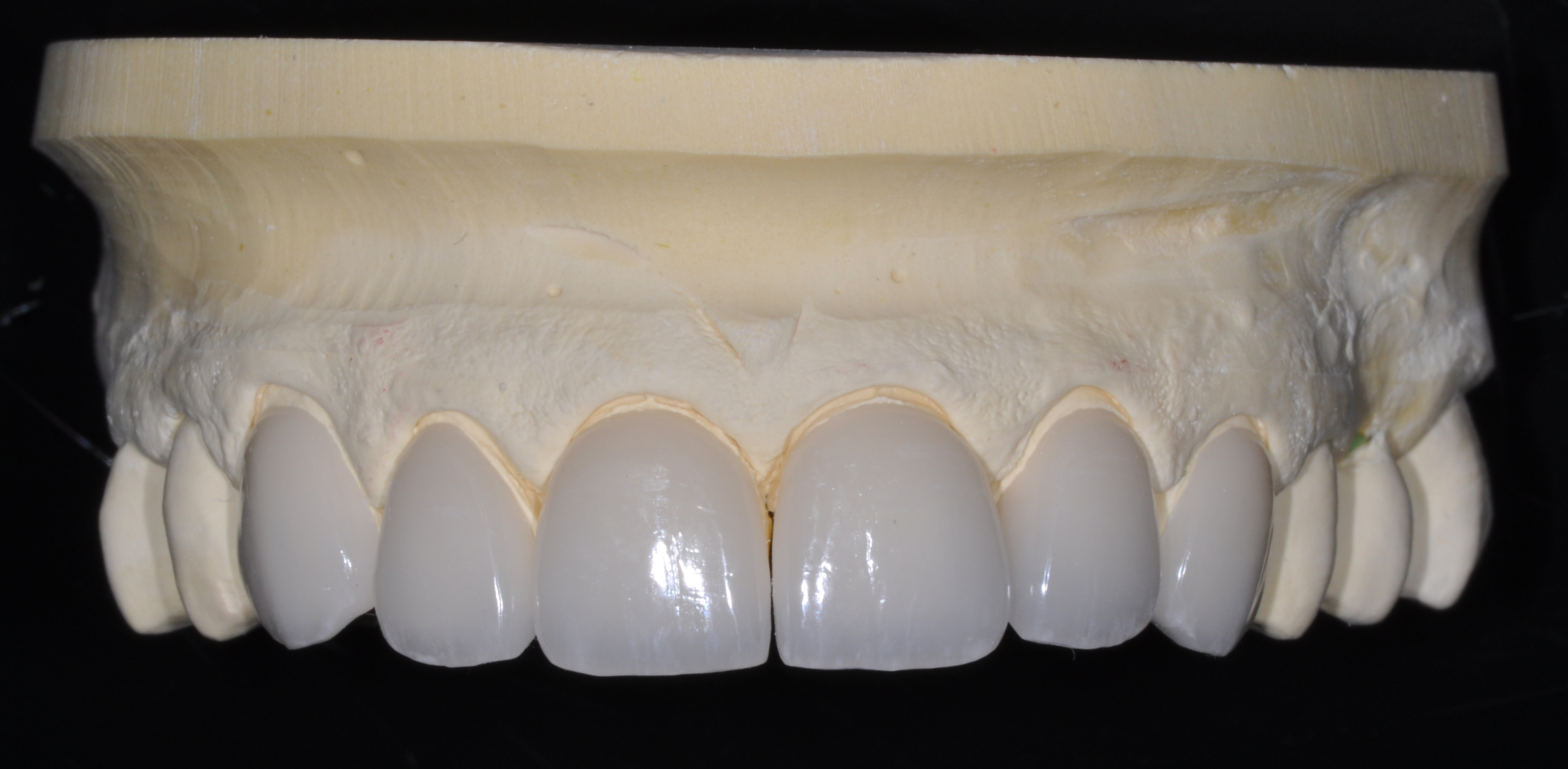
Feldspathic ceramic veneers.
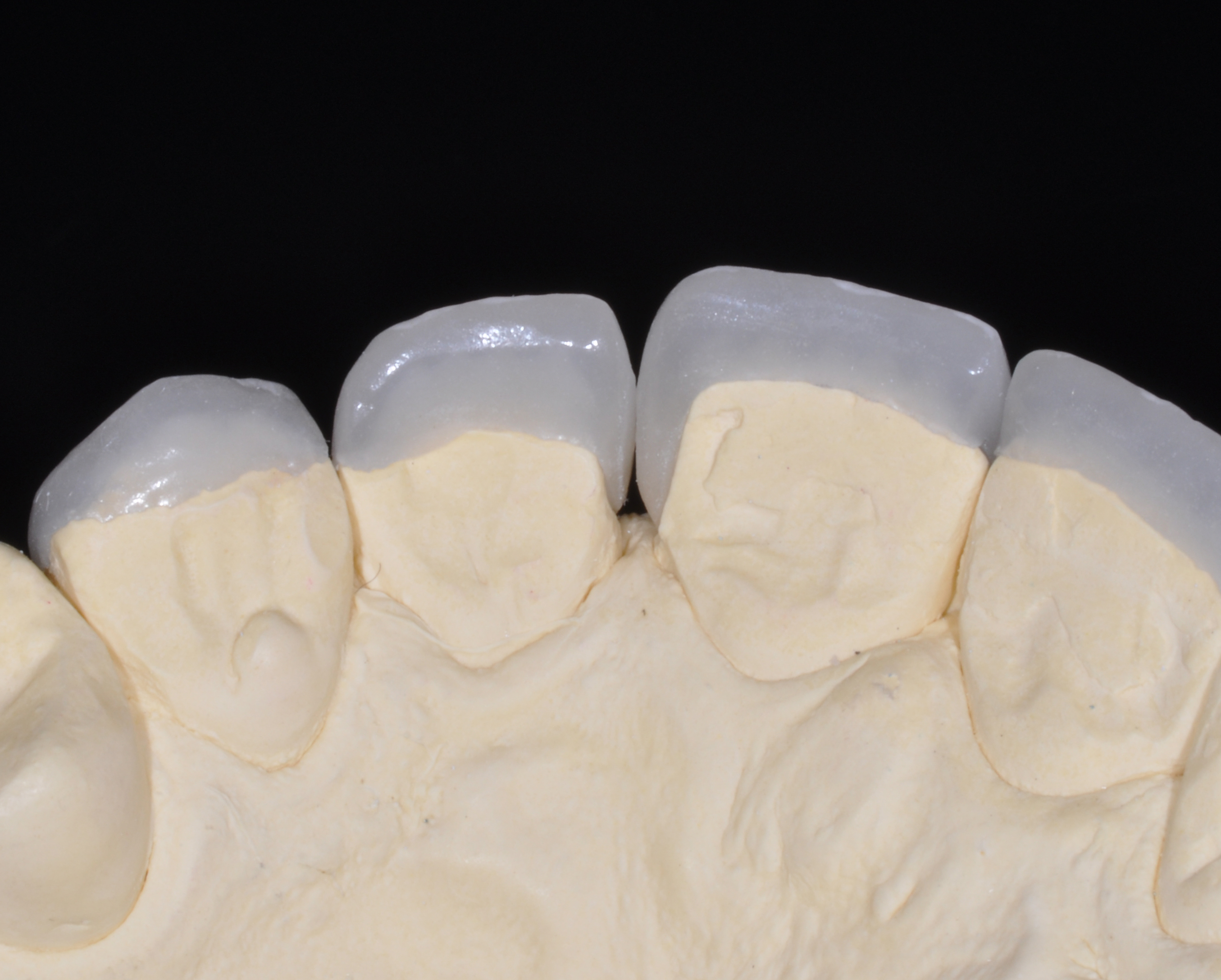
Type IV plaster master model showing perfect adaptation of the veneer in the palatal area.
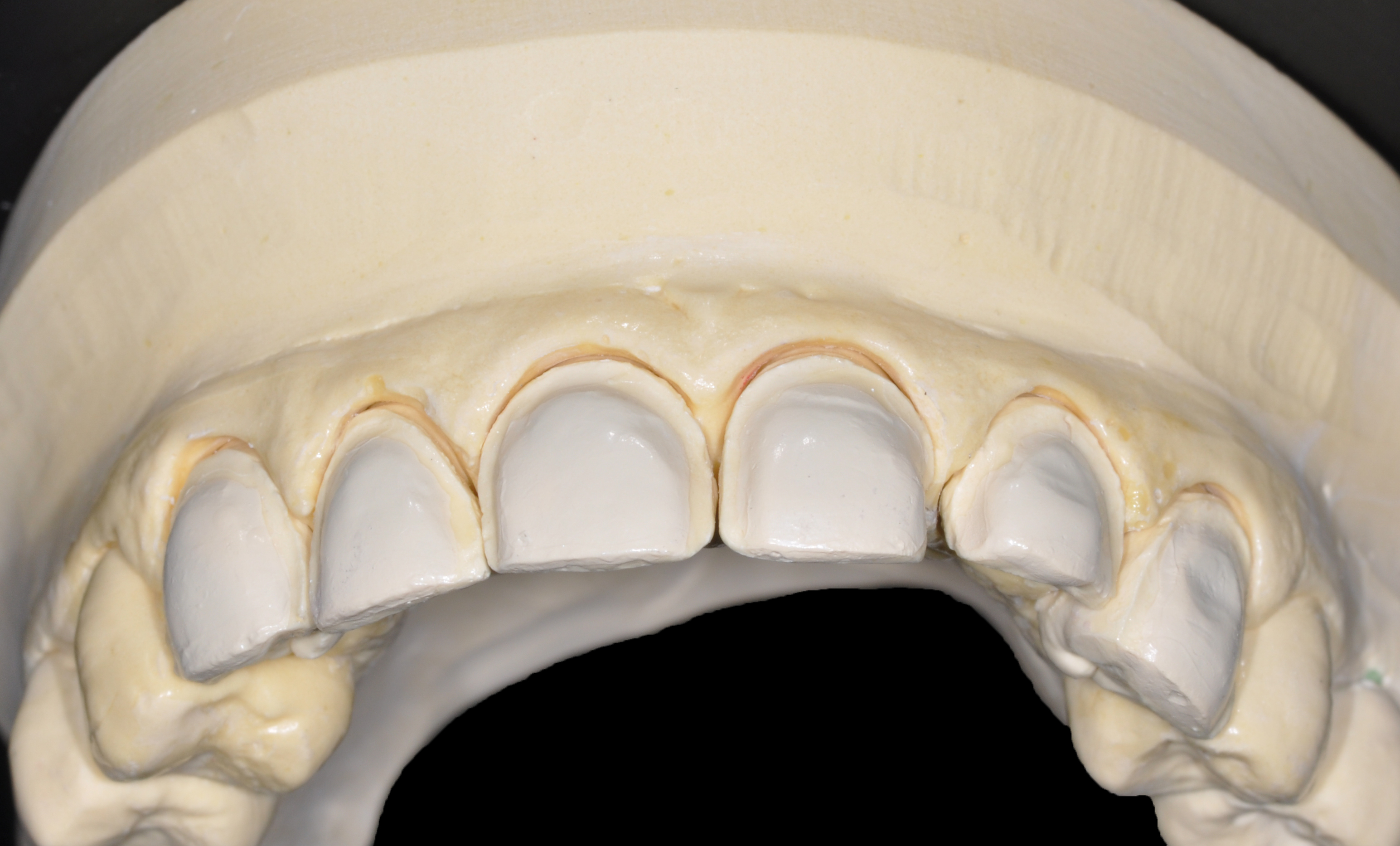
Close-up of the master model made with Type IV plaster.
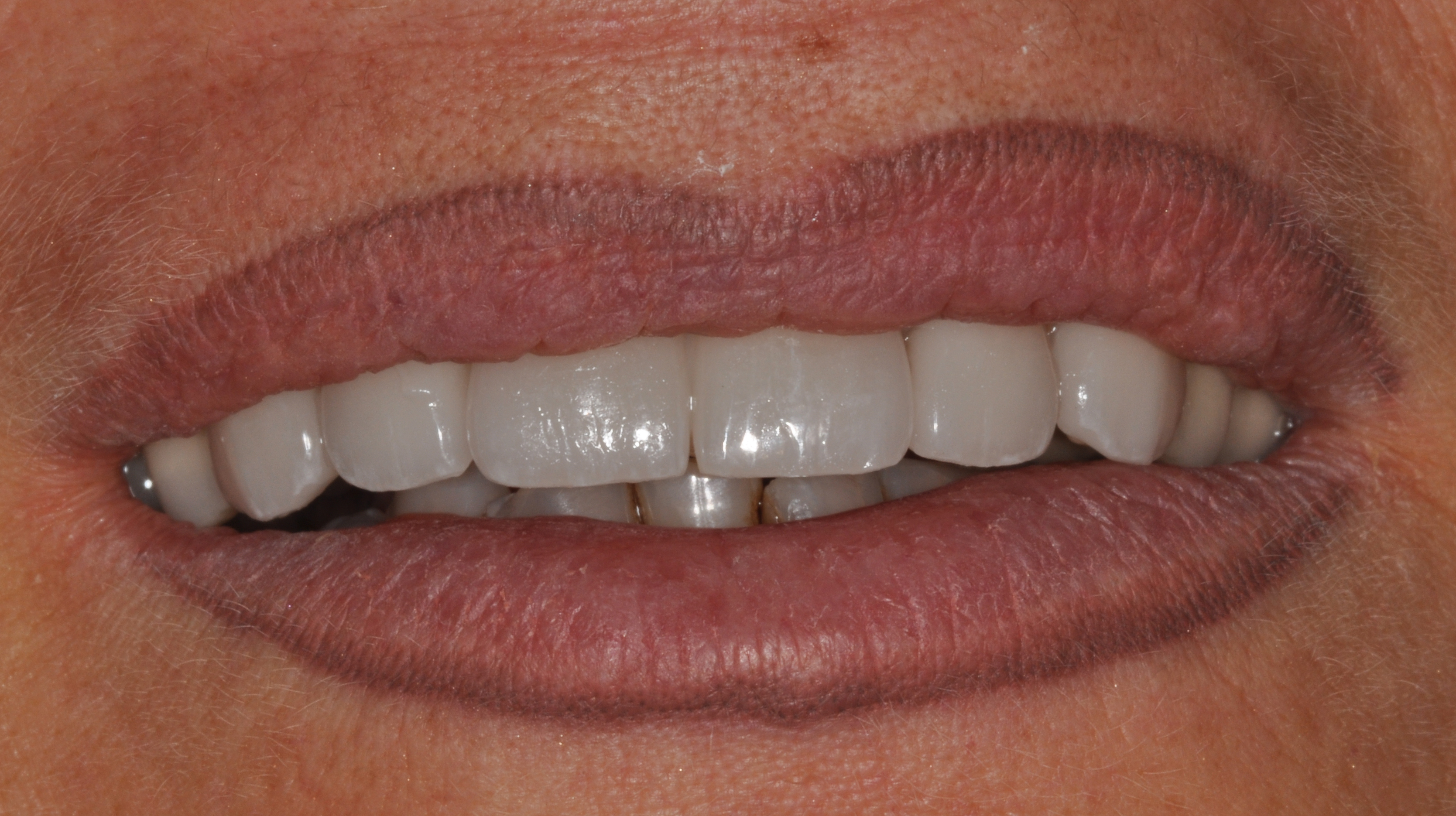
Application of veneers with try-in gel (NX3 Kit®, Kerr Dental).
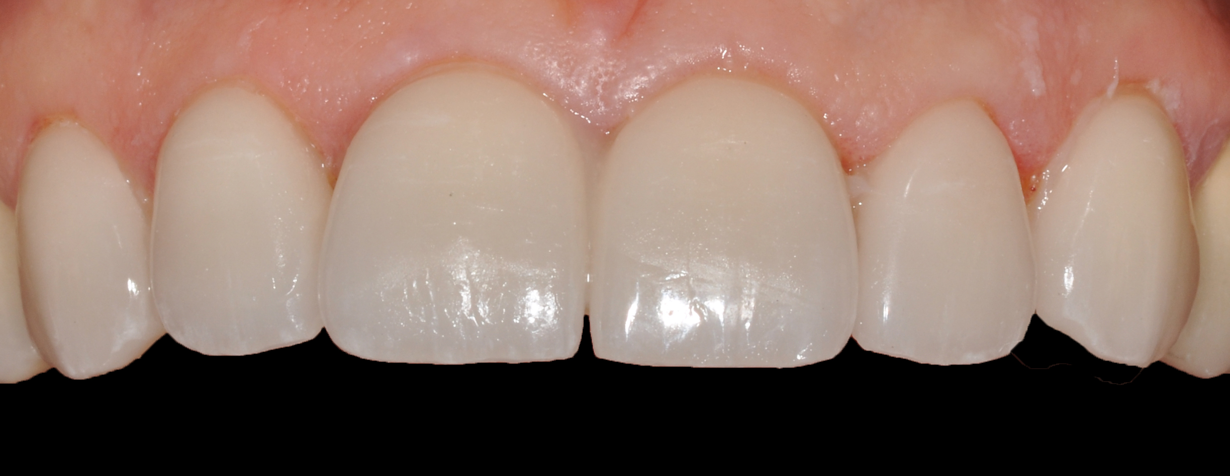
Close-up of veneers with try-in gel (NX3 Kit®, Kerr Dental).
2.8. Bonding Protocol
As silica-based restorations, the internal surfaces of the veneers must be etched with 9.5% hydrofluoric acid (HF) for 20 seconds (lithium disilicate) or 60 seconds (feldspathic porcelain). After thorough rinsing, the veneers should be placed in an ultrasonic bath (distilled water, 95% alcohol, or acetone) for 4 minutes to remove residual salts. Once dried, a silane primer is applied for 1 minute to facilitate a chemical covalent bond, followed by gentle air-drying to evaporate the solvent. The use of a rubber dam is mandatory for adequate isolation (Figs. 16, 17).
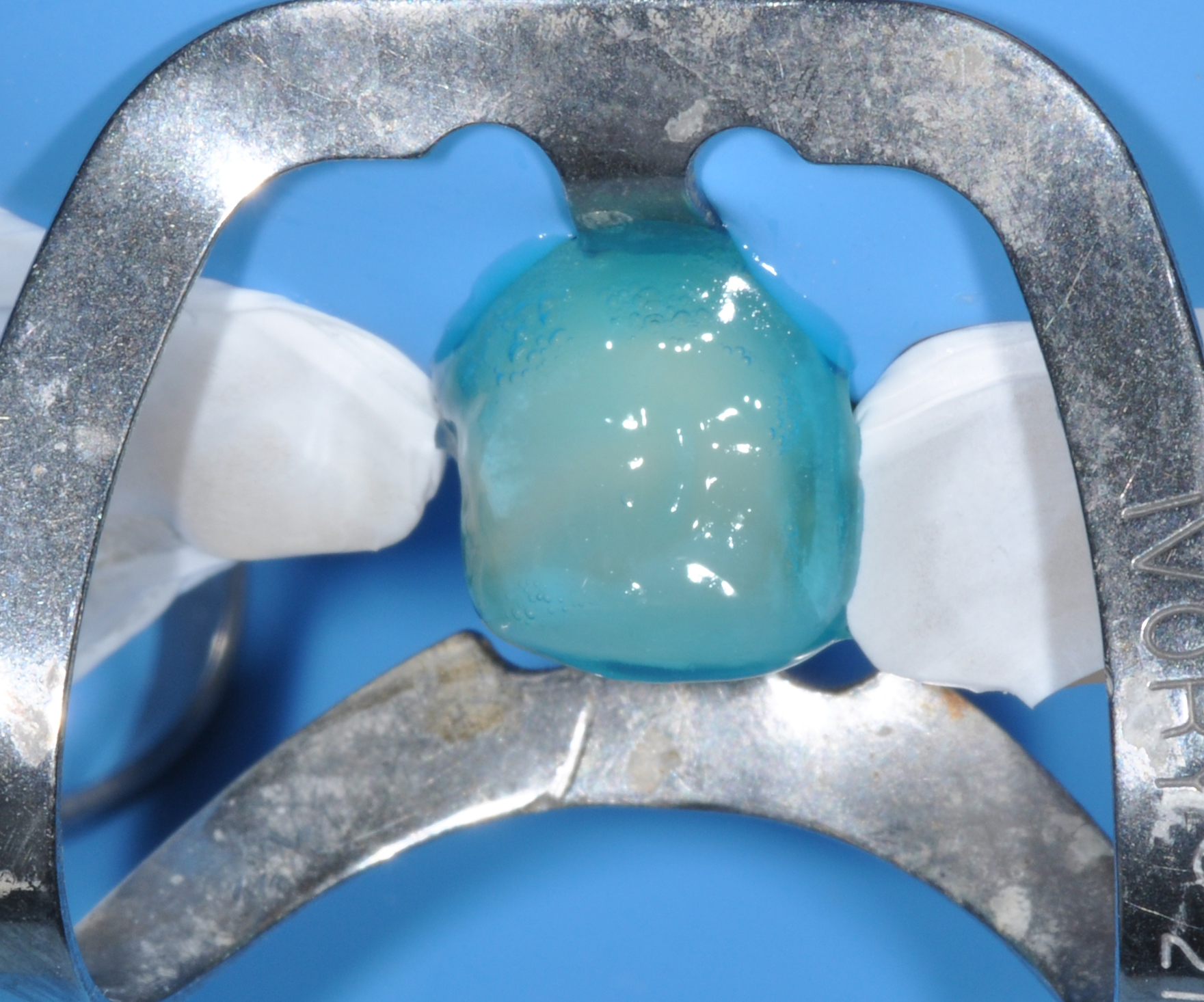
Single-tooth etching technique after dental dam isolation.
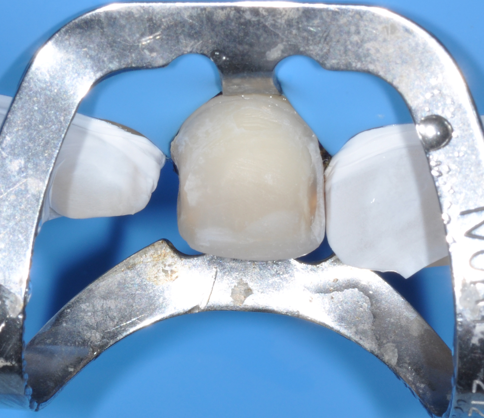
Tooth 1.1 showing typical etched enamel.
Light-cured composite resins are preferred for cementation due to their extended working time compared to dual-cure systems (Fig. 18). This allows for thorough removal of excess resin prior to polymerization, minimizing finishing time. The color of the resin cement typically accounts for approximately 10% of the final aesthetic outcome [1-5, 11, 12].
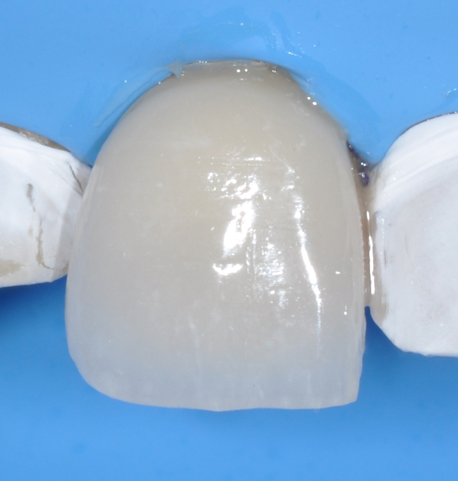
Veneer cementation on tooth 1.1 (NX3® photocuring cement, Kerr Dental).
2.9. Finishing and Polishing
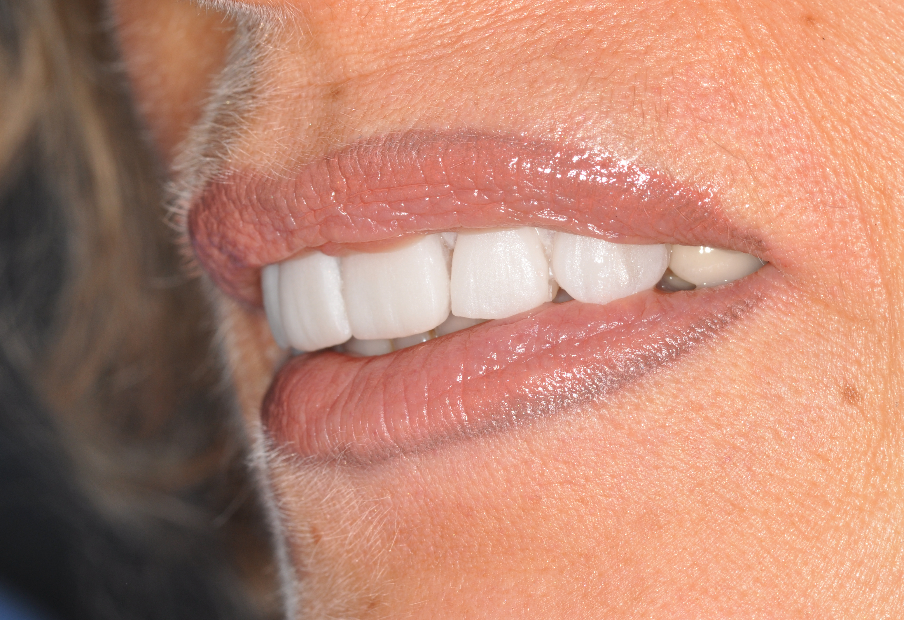
When bonding is properly executed, only minimal excess cement removal is required (Fig. 19). A #12 surgical blade is often preferred for precise marginal cleanup, as it reduces the risk of damaging restoration margins compared to rotary instruments. If further refinement is necessary, fine-grit diamond burs may be used, followed by thorough polishing to restore surface luster. Occlusion should be carefully evaluated in both centric and dynamic movements. Any adjustments performed with rotary instruments must be followed by re-polishing to maintain surface integrity (Fig. 20–23). Ultimately, the longevity and clinical performance of porcelain veneers (Fig. 24) rely on appropriate case selection, comprehensive diagnosis, and strict adherence to adhesive protocols
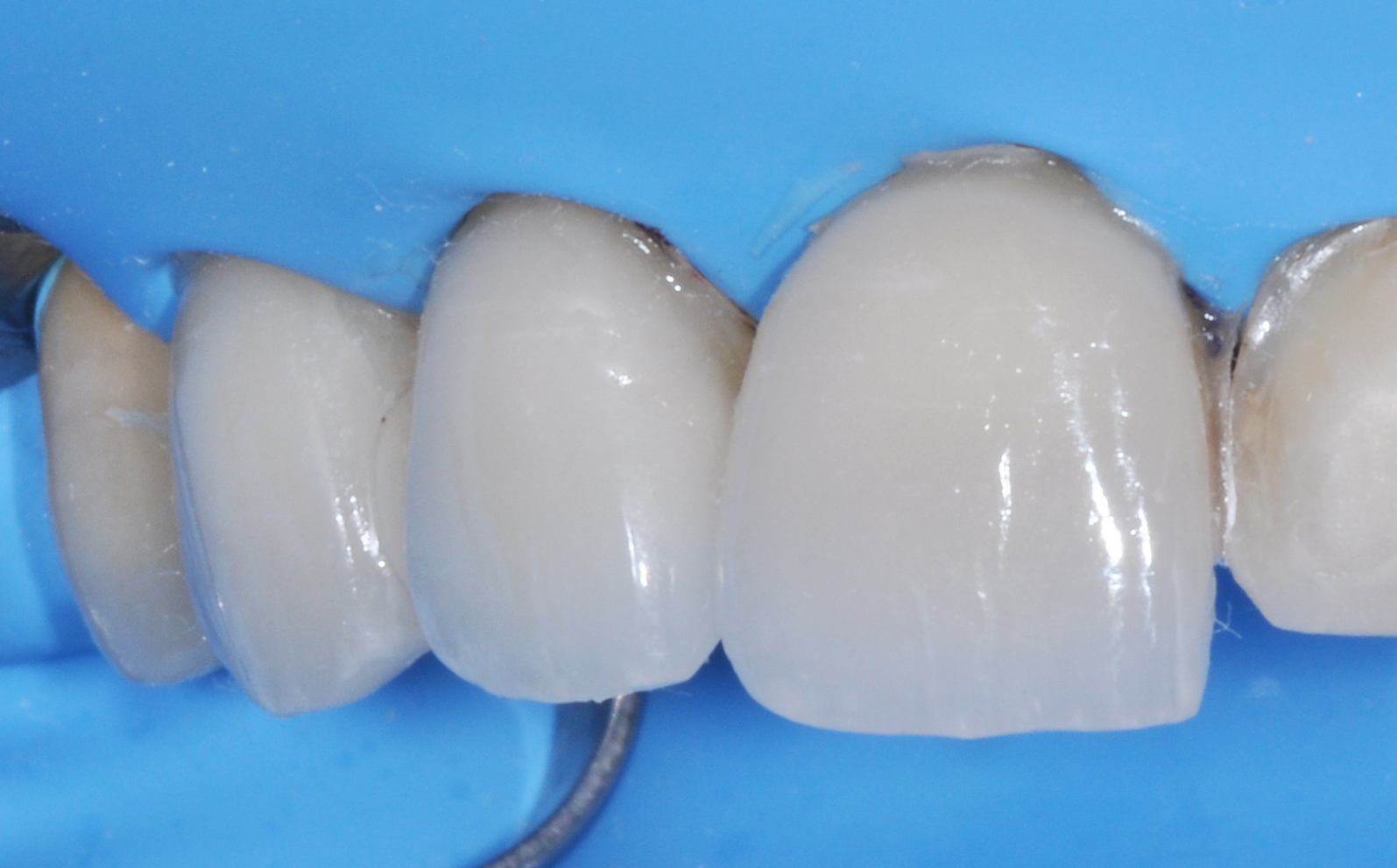
All veneers luted on first quadrant.
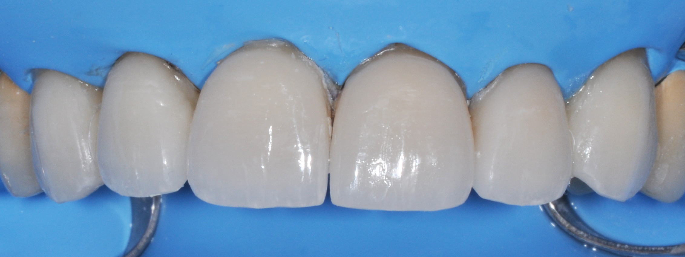
Veneers cemented on all upper jaw.
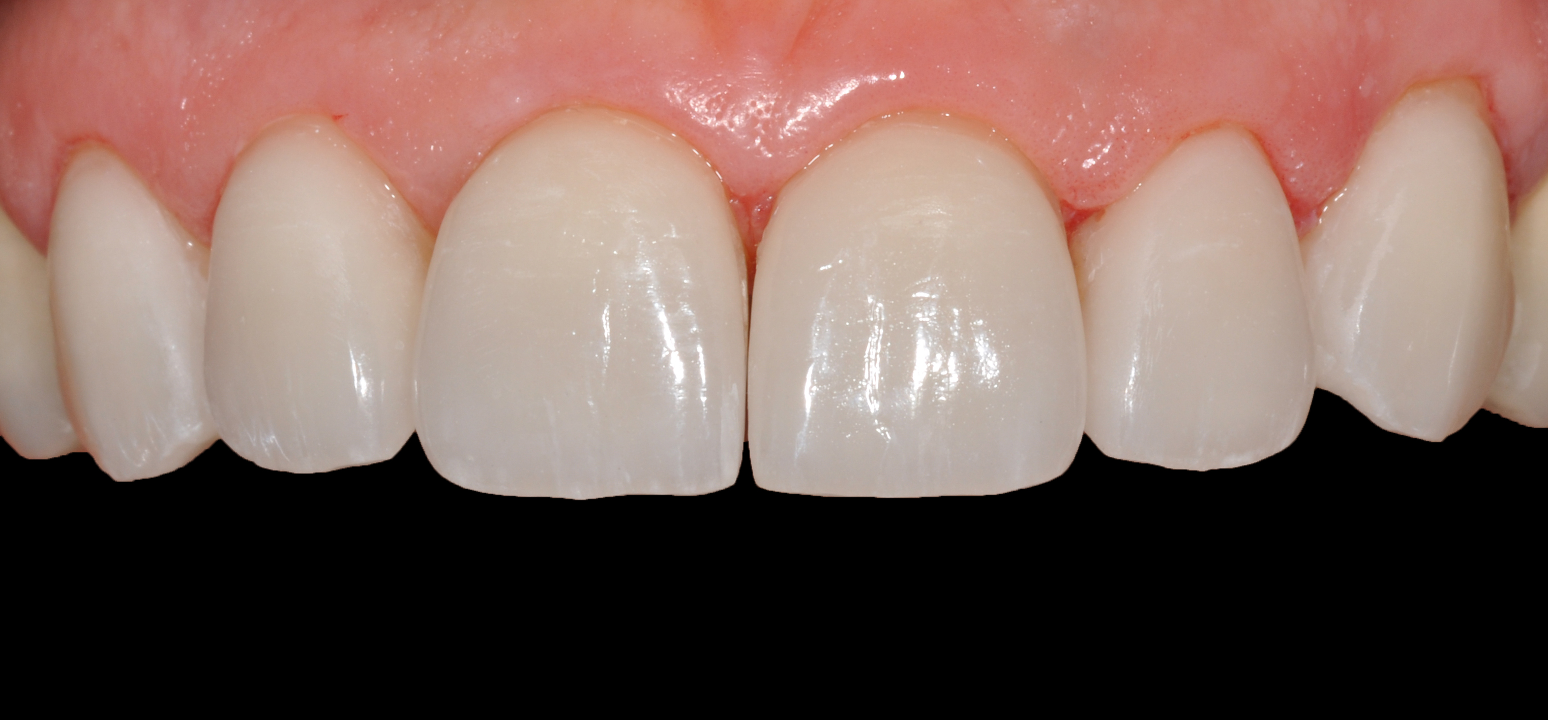
Post-op image showing all veneers luted.
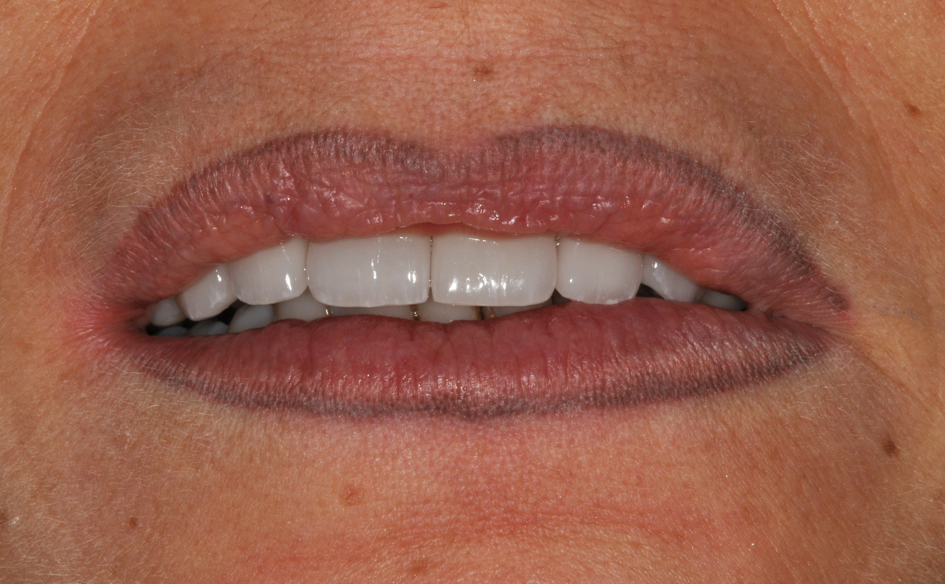
Immediate frontal post-op.
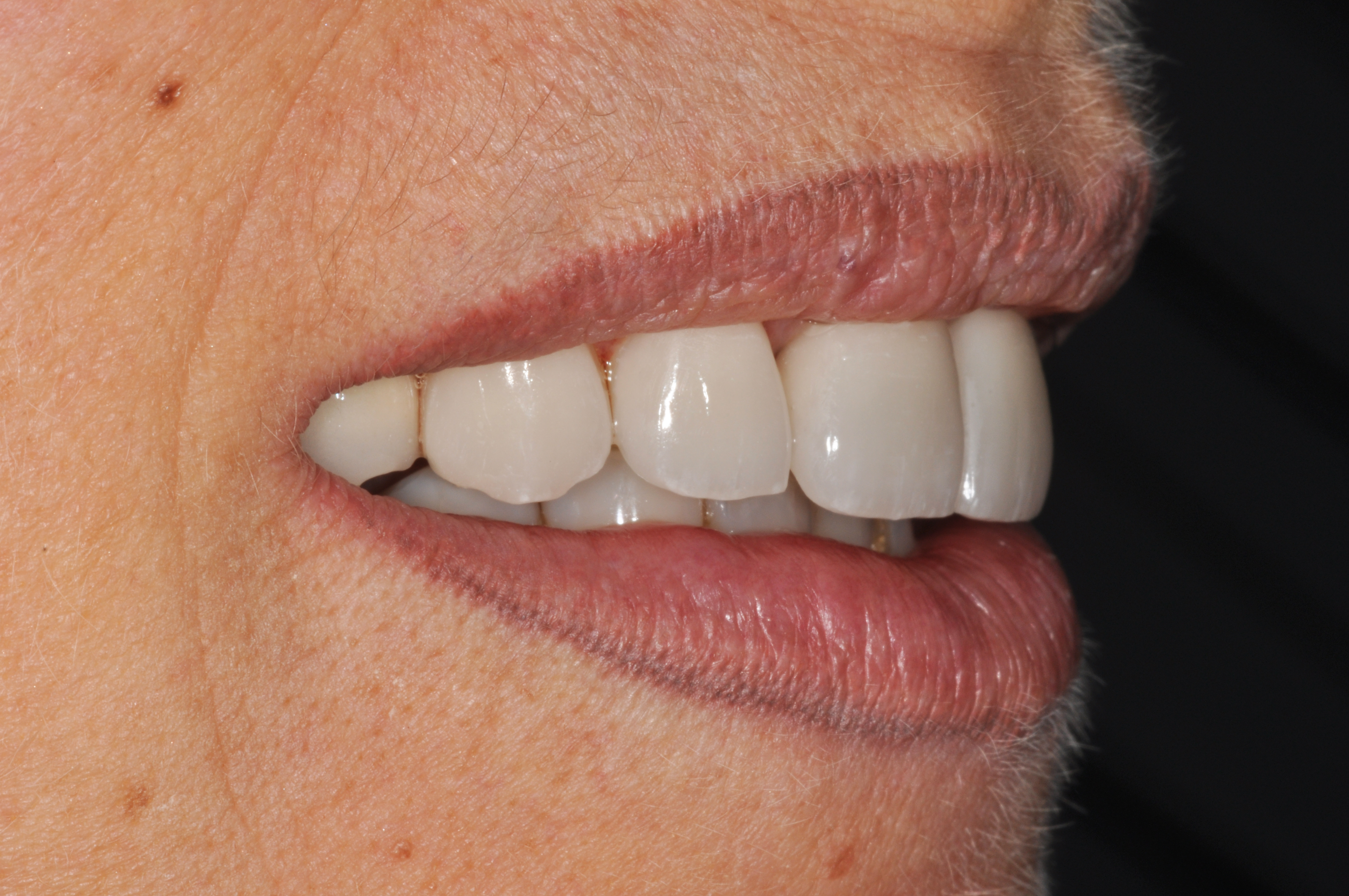
Immediate lateral post-op.
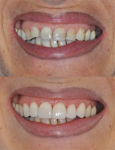
Comparison between Pre- and Post-op.
3. DISCUSSION
This case report describes a conservative and biomimetic approach for aesthetic rehabilitation with ceramic veneers, focusing on treatment planning, material selection, and clinical execution. Although ceramic veneers are well established in restorative dentistry, this report is clinically relevant because it addresses the replacement of aging direct composite restorations rather than the treatment of untreated teeth or severely compromised dentition. This condition is frequently encountered in daily clinical practice but is less commonly emphasized in the literature, where reports often focus on more complex or idealized situations. Direct composite restorations provide satisfactory short- and medium-term results; however, over time they are susceptible to discoloration, marginal deterioration, and surface wear, which may compromise aesthetics and require replacement with indirect restorations [6-9, 20, 21]. Preservation of enamel was a major objective in this case because long-term success of ceramic veneers is strongly associated with enamel bonding. Adhesion to enamel is more predictable and durable than adhesion to dentin because of enamel’s more favorable structure and lower permeability [1-5]. Since sufficient enamel was present, a minimally invasive preparation design was possible, in accordance with biomimetic restorative principles [22]. In addition, the patient’s age required careful consideration of gingival architecture, smile dynamics, and optical integration with adjacent teeth, confirming the importance of individualized treatment planning. Another important aspect of this case was the use of a guided and patient-centered workflow. Diagnostic wax-up, silicone reduction guides, and intraoral mock-up were not used only for visualization, but also as tools for treatment validation before irreversible preparation. The mock-up allowed assessment of tooth proportions, smile line, and patient expectations, improving communication among clinician, technician, and patient. This protocol reduced uncertainty and increased patient confidence in the final result. Previous studies have emphasized that mock-up procedures represent an essential step in aesthetic dentistry because they improve predictability and support shared decision-making [12-14, 20]. The development of ceramic veneers has evolved considerably since their first description by El-Mowafy [10]. Advances in adhesive dentistry, particularly acid-etching techniques and resin bonding protocols, have transformed veneers into reliable long-term restorations [1, 2, 11-13]. In the present case, strict adherence to adhesive procedures was fundamental. Hydrofluoric acid etching of the ceramic surface, silane application, enamel conditioning, and adhesive resin cementation were performed carefully to optimize bond strength and long-term stability. Material selection was also decisive for the final aesthetic result. Feldspathic porcelain was chosen because of its superior translucency and its ability to reproduce the optical behavior of natural enamel. Although lithium disilicate offers greater mechanical resistance, feldspathic ceramics remain highly indicated when maximum aesthetic integration is the primary objective. Clinical studies have shown high survival rates of ceramic veneers when preparation design is conservative and bonding protocols are properly followed [12, 20, 21]. The use of a light-cured resin cement also contributed to color stability and allowed better control of working time during cementation, both of which are important for highly aesthetic anterior restorations [18]. The color and condition of the underlying substrate were also considered, since pre-existing restorative materials and tooth discoloration may influence the final optical outcome [19]. The final result showed harmonious integration with the surrounding dentition and soft tissues, as well as high patient satisfaction. Functional analysis confirmed stable occlusal relationships, which are essential for the longevity of ceramic restorations and for minimizing the risk of fracture or debonding. These findings are consistent with previous reports indicating that successful veneer treatment depends on accurate case selection, conservative preparation, and strict compliance with adhesive protocols [12, 13, 20]. This case reinforces the importance of preserving enamel, using pre-operative visualization, and following meticulous adhesive procedures to obtain predictable and reproducible aesthetic outcomes. It also highlights that successful treatment is not determined only by technical execution, but also by effective communication and alignment with patient expectations. A patient-centered approach remains essential for achieving both functional success and long-term aesthetic satisfaction.
CONCLUSION
In conclusion, successful ceramic veneer treatment depends on a balance between conservative tooth preparation, accurate planning, and precise clinical execution. The preservation of enamel remains essential for achieving durable adhesion and long-term stability. Diagnostic wax-ups and intraoral mock-ups are valuable tools for evaluating aesthetics, guiding preparation, and improving communication with the patient before irreversible procedures are performed. When proper material selection and adhesive protocols are followed, ceramic veneers can provide stable functional performance together with natural and harmonious aesthetic results. The integration of digital tools and teledentistry may also contribute to treatment planning, professional education, and the clinical management of aesthetic restorative procedures.
AUTHOR’S CONTRIBUTION
The authors confirm contribution to the paper as follows: M.M.: Study conception and design; M.M.: Data collection; M.M.: Analysis and interpretation of results; M.M.: Draft manuscript. The author reviewed the results and approved the final version of the manuscript.
LIST OF ABBREVIATIONS
| MTA | = Mineral Trioxide Aggregate |
| HF | = Hydrofluoric acid |
| NiTi | = Nichel-titanium |
AVAILABILITY OF DATA AND MATERIALS
All the data and supporting information is provided within the article.
ACKNOWLEDGEMENTS
Declared none.