All published articles of this journal are available on ScienceDirect.

Management of Severely Angulated Implants using Both Angled Abutments and Mesostructures: A Clinical Report

Abstract
Background
Rehabilitation of the edentulous maxilla with a pneumatized sinus can be achieved through various approaches, including sinus lift procedures, short implants, and angulated implants. Angulated implants offer advantages by reducing cost, minimizing cantilevers, and avoiding extensive grafting. Optimal angulations (15°–45°) should be planned using cone beam computed tomography, and a surgical guide is recommended to ensure proper implant positioning for full-arch screw-retained prostheses.
Case Presentation
55 y old Caucasian female presented to Prosthodontic clinic at New Jersey Dental School with a chief complaint of restoring implants with a fixed prosthesis. Upon examination, the patient had 6 maxillary implants in place with the posterior implants at severe distal angle bilaterally, and the anterior implants at a severe labial angle. An unfavorable angulation between implants remained, even after correction with 30-degree multiunit abutments. Surveying the master cast showed that a single path of insertion between all abutments was not possible. Therefore, 3 separate mesostructures were fabricated to correct the remaining angulation problems, and a prosthesis superstructure was fabricated. Threads were tapped into each mesostructure, allowing the superstructure prosthesis to be screw retained. The patient was pleased with the function, comfort, and esthetics of her new prosthesis.
Conclusion
Angulated implants can successfully support full-arch fixed prostheses when planned with cone beam computed tomography and guided surgery. When a single insertion path is unachievable, separate mesostructures and a superstructure can help in screw-retained rehabilitation.
1. INTRODUCTION
Several treatment modalities for the edentulous maxilla with a pneumatized maxillary sinus have been introduced. Among these modalities are external sinus lift, short implants, and angulated implants [1]. Angulated implants offer several advantages, including reduced treatment costs, minimization of the cantilever, avoidance of extensive sinus grafting procedures, and consequently, shorter treatment times and reduced postoperative morbidity [2-4]. The recommended angulation of the distal implants should range between 15° and 45° to allow correction with multi-unit abutments. Treatment planning using Cone-Beam Computed Tomography (CBCT) before surgery is crucial for the success of full-arch rehabilitation. The implant angulations should be determined using planning software, and a surgical guide should be used during surgery to ensure a favorable path of insertion for a screw-retained, full-arch, implant-supported prosthesis [5]. This clinical report presents a detailed technique to restore full-arch severely angulated maxillary implants with a screw-retained prosthesis using mesostructure and superstructure.
A 55-year-old Caucasian female presented to the Prosthodontic clinic at New Jersey Dental School with a chief complaint of restoring implants with a fixed prosthesis. The patient's medical history was reviewed and was not significant. Dental history revealed that she underwent maxillary full-arch extractions due to caries and a complete denture five years ago. She was not comfortable with the removable complete denture, so she had six implants in the maxilla to support a full-arch fixed prosthesis one year ago. The distal implants were tilted to avoid an external sinus lift. In the mandible, she has a single crown on tooth # 38 and multiple splinted surveyed crowns from canine to canine. A Removable Partial Denture (RPD) was performed 5 years ago to replace the missing mandibular teeth. The patient was not comfortable with the RPD, so she had 3 restored implants in quadrant four two years ago and three unrestored implants 1 year ago. Her oral hygiene was fair, and she reported that she brushes her teeth twice a day without using dental floss.
Clinical examination revealed no muscle tenderness, palpable lymph nodes, or signs of temporomandibular disorders. The soft tissues of the lips, cheeks, tongue, hard palate, and soft palate were within normal limits. The gingival color and texture were also within normal limits. The probing depths of the remaining mandibular teeth ranged from 1 to 3 mm, with localized bleeding on probing. The width of the attached gingiva ranged from 1 to 6 mm around the implants and from 2 to 5 mm around the remaining mandibular teeth. In the maxillary arch, six implants were placed at positions 16, 13, 11, 21, and 27 using a narrow platform (3.5 mm in diameter) and at position 24 using a regular platform (4.3 mm in diameter) (Replace Select; Nobel Biocare, Göteborg, Sweden). Healing abutments were placed on implants #13, 11, 21, and 24, while 30° multiunit abutments were placed on implants #16 and 27. In the mandible, there was a single Porcelain-Fused-To-Metal (PFM) surveyed crown on tooth #38 and 6 units of splinted PFM crowns from 33-43. Also, there were 3 unrestored implants #34, 35, and 37 with healing abutments and 3 restored implants #44, 45, and 46 with PFM splinted crowns (Fig. 1).
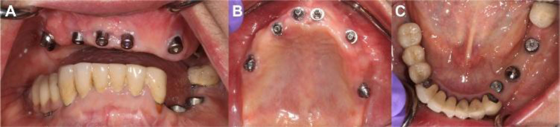
A: Frontal view. B: Maxillary occlusal view after placement of multiunit abutments. C: Mandibular occlusal view.
The panoramic radiograph showed normal bone trabeculation. The maxillary sinuses were severely pneumatized bilaterally. Mandibular inferior border and condyles were within normal limits. Six implants were placed in the maxilla, and the distal implants were severely angulated. (Fig. 2) Periapical radiographs showed good marginal adaptation of the surveyed crowns and periapical radiolucency related to tooth # 43.
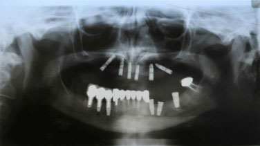
Panoramic radiograph.
The treatment plan, which included the construction of a full-arch, screw-retained, maxillary fixed prosthesis and a four-unit, implant-supported fixed dental prosthesis extending from positions 34 to 37 in the mandible, was approved by the patient. 30° multiunit abutments (Nobel Biocare, Göteborg, Sweden) were selected according to the cuff height and placed on implants #13, 11, 21, and 24 with the best possible angulation correction. Open-tray abutment-level impression for the maxillary implants and fixture-level impression for the mandibular implants were taken and poured to fabricate the master cast (Fig. 3).
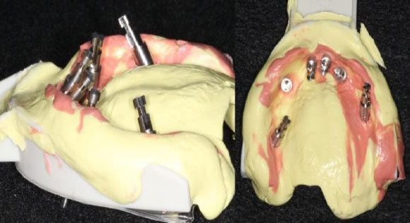
Maxillary final impression.
Jaw relation was performed in centric relation to mount the master cast, followed by teeth setup (Fig. 4).
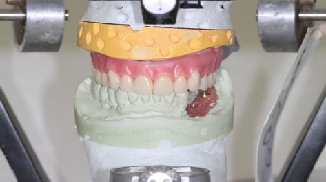
Mounted maxillary master cast with teeth set up.
Teeth try-in was verified and approved by the patient. A putty index was made to indicate the teeth position and evaluate the space available for the metal framework (Fig. 5).
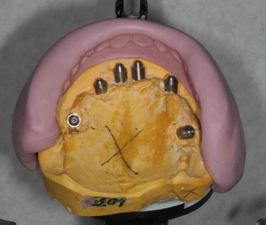
Maxillary master cast with teeth index.
The master cast survey showed no single possible path of insertion to fabricate a one-piece full-arch metal framework on all maxillary implants due to the severe angulation of the posterior and anterior implants (Fig. 6).
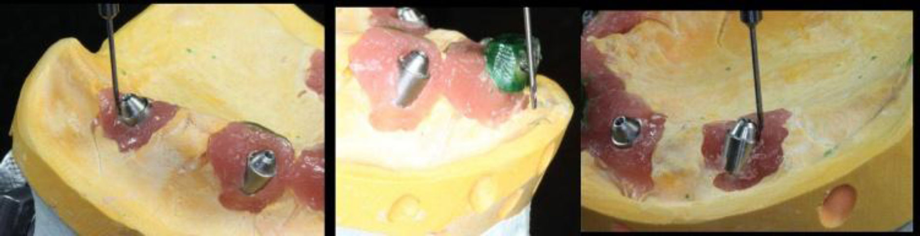
Surveying the maxillary master cast for a common path of insertion.
Instead of a single full-arch metal framework, three separate mesostructures were waxed and cast using a nickel-chromium (Ni-Cr) metal alloy (Wiron 99; Bego, Bremen, Germany). Each mesostructure connected two implants (16–13, 11–21, and 24–27) and was designed with parallel side walls to establish a single path of insertion for the superstructure. The separate mesostructures were tried in the patient's mouth to ensure a passive fit using visual inspection, radiographic examination, and the one-screw test [5, 6]. Cutting and soldering were required in the posterior segments. Therefore, the mesostructures were sectioned and joined intraorally using acrylic resin (GC Corp., Tokyo, Japan) and then sent to the laboratory for soldering (Fig. 7).
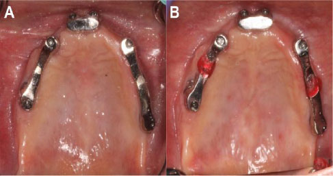
A: Separate mesostructures try-in. B: Sectioning and joining the posterior mesostructures for the passive fit.
After soldering, the mesostructures were tried in the patient's mouth, and passive fit was achieved. Interocclusal record at the proposed vertical dimension in centric relation was taken. An open tray pickup impression using long impression coping pins was taken, then used to fabricate a new master cast (Figs. 8 and 9).
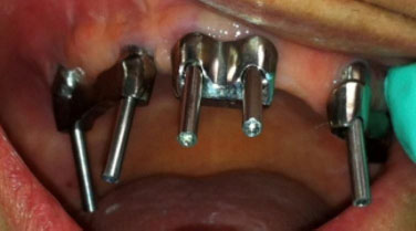
Mesostructures pick-up impression after cut and solder.
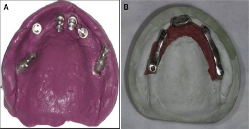
A: New pick-up final impression. B: New maxillary master cast with mesostructures after cut and solder.
After mounting the new master cast, a putty index was fabricated over the maxillary and mandibular teeth on the old master cast to transfer the tooth setup to the new master cast (Figs. 10–12).
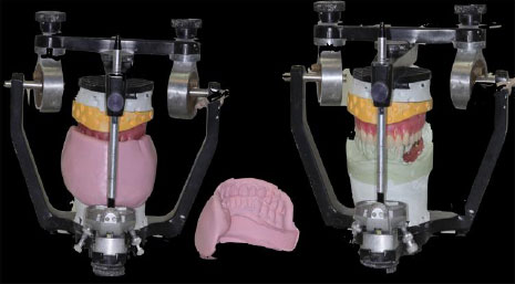
Putty index to transfer the teeth position from old master cast to the new one.
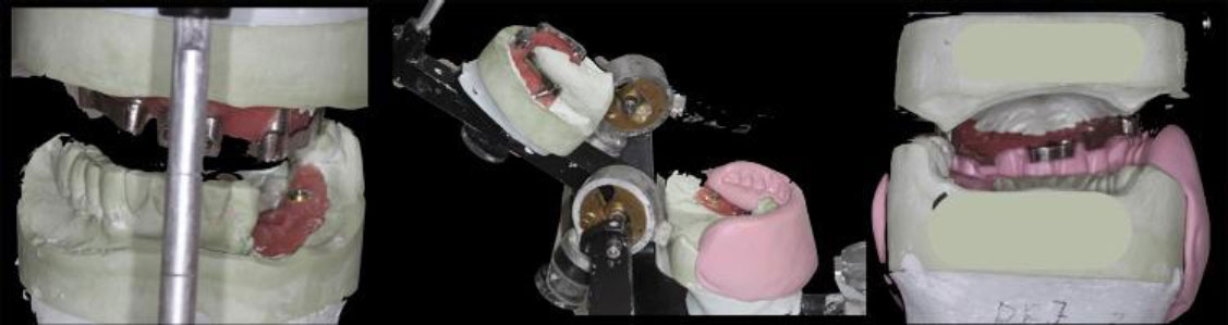
Mounted new master cast with teeth index.
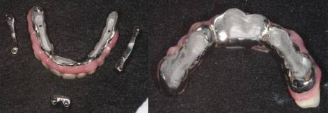
Screw-retained final prosthesis after tapping threads in the mesostructured.
Full arch superstructure was then waxed and cast using nickel-chromium (Ni-Cr) metal alloy (Wiron 99, Bego, Bremen, Germany) to fit over the mesostructure. The tooth setup was performed using the index. Three threads were tapped in the mesostructure to allow the full-arch superstructure to be screw-retained. The waxed final prosthesis was tried in the patient's mouth, and the esthetics, phonetics, and group-function occlusal scheme were verified. The final prosthesis was then processed into acrylic resin to fabricate the final hybrid prosthesis. The mandibular fixed dental prosthesis 34-37 was then fabricated and delivered with the maxillary full arch fixed prosthesis. Surveyed crown #43 was sectioned and removed to treat the periapical pathology. Assessment of the remaining tooth structure revealed that the tooth was non-restorable; therefore, it was extracted and planned to be replaced with a single implant-supported crown in the future. The patient was satisfied, and the esthetics and function were restored (Figs. 13 and 14).
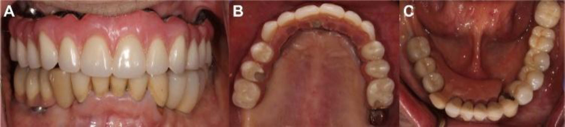
A: Final prosthesis frontal view. B: Final prosthesis maxillary view. C: Final prosthesis mandibular view.
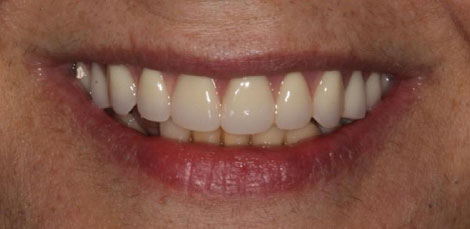
Final prosthesis smile.
A follow-up appointment was conducted one week later to evaluate and adjust the occlusion as necessary, and a night guard was provided to the patient. The patient will attend regular follow-up appointments every six months to evaluate the success of the rehabilitation protocol and detect any potential complications.
3. DISCUSSION
The main treatment modalities for edentulous patients are either fixed or removable prostheses [7]. Complete dentures are the most economical option; however, they have several disadvantages, including discomfort, poor denture stability, difficulty with eating, and inadequate or compromised retention [8]. Therefore, dental implants have been used either to improve the retention of removable dentures, such as overdentures, or to replace them with fixed prostheses [1]. A review of the literature noted that prostheses supported by dental implants significantly improved the quality of life for edentulous patients [8]. Rehabilitation of the edentulous maxilla with an angulated abutment was first introduced with the all-on-four concept to avoid sinus lifting and minimize cantilever length [9]. The main disadvantage of this concept is that a fixed prosthesis cannot be used if one of the implants fails. In this case, implants were placed using the same concept due to pneumatized sinuses, with 6 implants placed to allow the prosthesis to be used even if one implant failed and for improve biomechanical behavior [10]. Babacan et al. compared the biomechanical behavior between angled and vertical implants and showed that the angled implant demonstrated a higher stress value in the surrounding bone [11]. However, increasing the number of implants showed better stress distribution between all implants [11]. Implants placed with unfavorable angulations, exceeding 45° between the anterior and posterior implants, can be restored with either an overdenture or a cement-retained fixed prosthesis due to the lack of a common path of insertion for a screw-retained fixed prosthesis. There are several disadvantages associated with the use of cement-retained prostheses for full-arch rehabilitation, including the difficulty of removing excess cement, which may lead to serious biological complications around the implants [12]. Another disadvantage is the difficulty in retrievability of the prosthesis for maintenance when needed [12]. Therefore, a solution for managing severely angulated implants to be restored with a screw-retained prosthesis was used in this case. A hybrid prosthesis using metal-resin rather than a ceramometal prosthesis has several advantages and disadvantages. The advantages include reduced cost and ease of replacing the acrylic teeth in the future, if needed [13]. The main disadvantage is increased wear, particularly when the prosthesis is opposed by a ceramic restoration. In this case, the patient was provided with a night guard to minimize wear associated with parafunctional habits [14].
CONCLUSION
The use of angulated implants for the rehabilitation of edentulous arches with full-arch fixed prostheses can provide a predictable treatment option when comprehensive treatment planning is performed using Cone-Beam Computed Tomography (CBCT) and guided surgical protocols. However, in certain clinical situations, achieving a common path of insertion for the definitive full-arch prosthesis may remain challenging despite the use of multi-unit abutments to compensate for implant angulation. In such cases, the use of separate mesostructures supporting a unified superstructure may provide an effective alternative, facilitating the fabrication and delivery of a screw-retained full-arch prosthesis while accommodating the divergent implant axes.
AUTHOR’S CONTRIBUTION
The author confirms sole responsibility for the following: N.M.: Study conception and design, data collection, analysis and interpretation of results, and manuscript preparation.
LIST OF ABBREVIATIONS
| CBCT | = cone beam computed tomography |
| RPD | = removable partial denture |
| PFM | = porcelain fused to metal |
ETHICS APPROVAL AND CONSENT TO PARTICIPATE
The clinical records, radiographs, and laboratory data were obtained directly from the patient during routine care at New Jersey Dental School at Rutgurs University, in accordance with institutional ethical standards and with the patient's informed consent and approval from the institutional review board was not required.
HUMAN AND ANIMAL RIGHTS
All procedures involving human participants were conducted in accordance with the ethical standards of the committee responsible for human experimentation (institutional and national), and with the Helsinki Declaration of 1975, as revised in 2013.
AVAILABILITY OF DATA AND MATERIALS
The data supporting the findings of the article will be available from the corresponding author (N.M) upon reasonable request.
ACKNOWLEDGEMENTS
The author thanks Dr. LD, DMD, FACP, and Dr. RF, D.D.S., M.S for their valuable support and guidance throughout the treatment.