All published articles of this journal are available on ScienceDirect.

Investigation of the Cytotoxicity and Anti-inflammatory Properties of Glass Ionomer Cement containing Titanium Dioxide Nanoparticles
 Simin Sharifi4 , *
Elmira Jafari Navimipour1
Mohammad Esmaeel Ebrahimi Chaharom1
Fathemeh Dabaghi Tabriz1
Simin Sharifi4 , *
Elmira Jafari Navimipour1
Mohammad Esmaeel Ebrahimi Chaharom1
Fathemeh Dabaghi Tabriz1
Authors Info & Affiliations
Abstract
Introduction
Glass Ionomer Cements (GICs) modified with bioactive Nanoparticles (NPs) have emerged as promising materials for enhanced performance in restorative dentistry. This study aimed to evaluate the cytotoxicity and anti-inflammatory properties of a novel GIC formulation incorporating titanium dioxide (TiO2) nanoparticles.
Methods
In this in vitro investigation, TiO2 NPs were incorporated into conventional GIC powder (Fuji II) at a concentration of 1 wt%. Human Gingival Fibroblasts (HGFs) were cultured in Dulbecco’s Modified Eagle Medium (DMEM) supplemented with Fetal Bovine Serum (FBS) and antibiotics. Cytotoxicity was assessed using the MTT assay at 24, 48, and 72 hours following exposure to the modified and unmodified GIC. To evaluate anti-inflammatory effects, HGFs were treated with either conventional GIC or TiO2-modified GIC for 24 hours. Untreated cells served as the control group. Following treatment, all groups were stimulated with 45 µg/mL lipopolysaccharide (LPS) to induce an inflammatory response. The concentrations of IL-6 and TNF-α were quantified using sandwich ELISA kits. Statistical analysis was performed using one-way and two-way ANOVA followed by Tukey’s post hoc test, with significance set at p < 0.05.
Results
The incorporation of TiO2 NPs did not significantly alter the cytotoxicity of GIC (p > 0.05). Both modified and unmodified GICs exhibited cytotoxic effects within the first 24 hours, with cell viability ranging from 55% to 60%. No significant differences in cytotoxicity were observed across the 24, 48, and 72-hour time points (p > 0.05). LPS stimulation significantly elevated IL-6 and TNF-α levels in all groups (p < 0.001), confirming robust immune activation. Notably, conventional GIC further enhanced IL-6 production in the presence of LPS (p < 0.01). In contrast, TiO2-modified GIC significantly attenuated the expression of both cytokines under inflammatory conditions (p < 0.01). Under non-stimulated conditions, cytokine levels remained low and did not differ significantly from the control group.
Discussion
The findings suggest that the addition of low-dose TiO2 NPs does not exacerbate the cytotoxic effects of GIC and may confer anti-inflammatory benefits, thereby enhancing the biological performance of GICs in restorative applications.
Conclusion
The presence of TiO2 NPs significantly decreased the expression of proinflammatory cytokines, suggesting a potential immunomodulatory benefit.
1. INTRODUCTION
Glass Ionomer Cements (GICs) are extensively employed in clinical dentistry, serving as cavity bases, liners, restorative materials, and luting agents. Their broad functional applicability is attributable to the capacity for tailoring physicochemical properties through deliberate modifications in formulation parameters, including alterations in the chemical composition and adjustments to the powder-to-liquid ratio [1]. GICs demonstrate several clinically favorable attributes, notably their intrinsic chemical bonding to dental hard tissues, biocompatibility, and sustained fluoride ion release. Despite these advantages, their clinical performance is constrained by inherent material deficiencies, including limited mechanical strength, suboptimal wear resistance, elevated aqueous solubility, and inadequate antibacterial efficacy. These shortcomings can contribute to bacterial colonization, secondary caries, tooth fracture, and eventual restoration failure [2]. Notably, secondary caries remains the predominant cause of failure in GIC-based restorations [3]. To address these limitations, various strategies have been employed to enhance the performance of GICs, including the incorporation of additives such as resin, hydroxyapatite, reinforcing fibers, and nanoparticles (NPs) into the glass powder matrix. These modifications have yielded mixed results in terms of improving both mechanical and biological properties [4]. Among these approaches, the integration of NPs has garnered significant attention in dental materials research [5, 6]. NPs can occupy interstitial spaces within the cement matrix, functioning as reinforcing agents that enhance structural integrity and mechanical performance [7]. Titanium dioxide (TiO2) NPs have been introduced into GIC formulations due to their favorable attributes, including excellent biocompatibility, chemical stability, mechanical strength, and antimicrobial activity [8, 9]. Their incorporation has shown promise in reducing cariogenic biofilm formation and enhancing the antibacterial efficacy of GICs [10]. Given the direct contact of restorative materials with periodontal tissues, it is imperative that these materials exhibit minimal cytotoxicity and do not elicit adverse tissue responses [11]. Therefore, alongside mechanical optimization, thorough evaluation of biocompatibility is essential to ensure safe clinical application [12, 13]. The biocompatibility of GICs is influenced by the chemical constituents released during the setting reaction, as both the glass powder and polyacid liquid components may exert cytotoxic effects [4].
In studies assessing the cytotoxicity of conventional glass ionomer cements on mouse fibroblasts, human fibroblasts, and human dental pulp stem cells, outcomes have varied according to the type of cement, the cellular model, and the time frame considered [14-23]. In a study conducted by Duzyol et al., glass ionomer cement demonstrated anti-inflammatory activity, significantly reducing the levels of TNFα, IL-1, and IL-6 [16]. While prior research has investigated the anti-biofilm, antibacterial, and physical and mechanical characteristics of glass ionomer cements incorporating titanium oxide nanoparticles, their biological properties have not been evaluated [10, 24, 25].
In the present in vitro study, GIC modified with TiO2 NPs was synthesized and evaluated for cytotoxicity and anti-inflammatory properties. Cell viability and cytokine expression were analyzed to determine whether TiO2 incorporation enhances the immunological safety profile of GIC without compromising its biocompatibility.
The null hypothesis of this study was that incorporation of 1 wt% TiO2 nanoparticles into glass ionomer cement would not significantly affect cytotoxicity or inflammatory cytokine expression compared to unmodified conventional GIC.
Unlike previous studies focusing mainly on mechanical or antibacterial properties, this study specifically evaluates the immunomodulatory response of TiO2-modified GIC under LPS-induced inflammatory conditions using human gingival fibroblasts.
2. MATERIAL AND METHODS
This in vitro experimental study was conducted following approval from the Ethics Committee of Tabriz University of Medical Sciences, Iran (IR.TBZMED.VCR. REC.1403.065), approval date: 2024-05-20.
2.1. Specimen Preparation
The powder of self-cured restorative glass ionomer cement (GC Fuji II, GC Corporation, Tokyo, Japan) was manually mixed with 1% titanium dioxide nanoparticles (TiO2, Sigma-Aldrich, Massachusetts, USA, particle size of 20 nm, Fig. 1). All materials were equilibrated to room temperature (22–25°C) prior to weighing and were handled quickly to minimize moisture uptake. The TiO2 NPs and GIC powder were kept in tightly sealed containers and opened only during weighing/mixing. To ensure accuracy in the process, both the cement powder and nanoparticles were carefully weighed using a digital scale (AND, HR20, UK) with a precision of 0.0001 g. Each component was weighed separately in pre-tared, clean weighing containers. The specified weight proportions of each component were then combined using a spatula (Royaniran, Tehran, Iran), and the mixing was continued manually until a homogeneous mixture was obtained. To reduce nanoparticle agglomeration, TiO2 NPs were incorporated gradually into the GIC powder while spatulating with consistent, standardized strokes; mixing was continued until no visible speckling or clumps remained. GIC containing 0 wt% TiO2 NPs was employed as the control group in this study. The control powder underwent the same handling and mixing procedure (excluding TiO2 addition) to ensure identical processing conditions across groups.
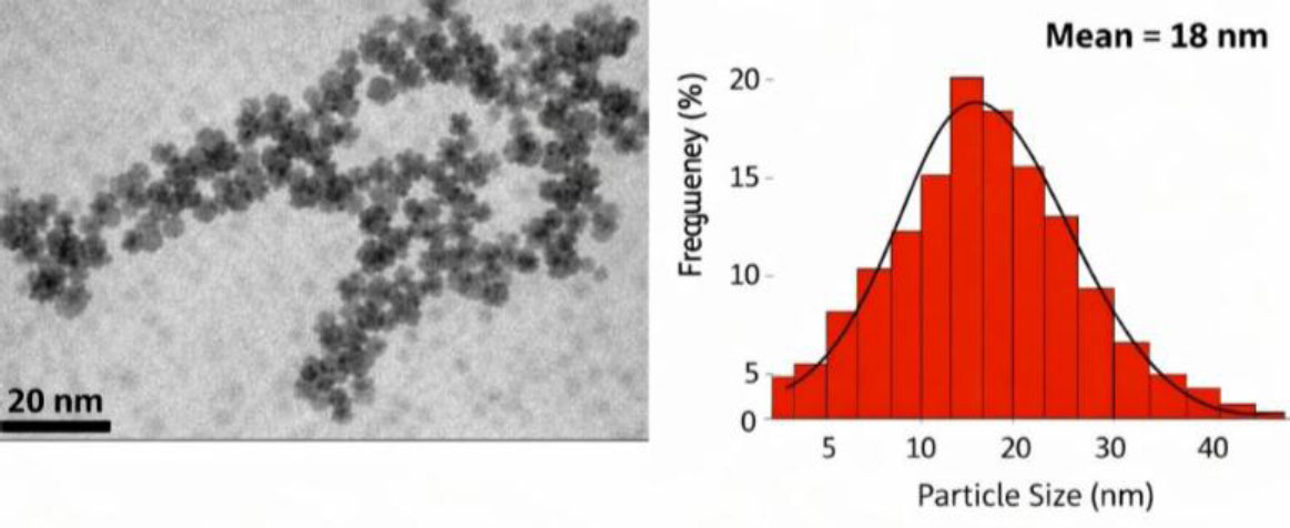
TEM image and mean particle size of the TiO2 NPs.
2.2. Cytotoxicity Evaluation
GIC disks containing 0 and 1 wt% TiO2 NPs were sterilized under ultraviolet (UV) light for 30 minutes. Each disk was immersed in serum-free Dulbecco’s Modified Eagle Medium (DMEM) at a ratio of 0.1 g/mL and incubated at 37°C with gentle agitation (50 rpm) for 24 hours to generate conditioned extracts. The extracts were subsequently filtered through 0.22 µm syringe filters, warmed to 37°C, and used immediately for cytotoxicity assays.
Human Gingival Fibroblasts (HGFs) were obtained from the Pasteur Institute of Iran and cultured in DMEM supplemented with 10% Fetal Bovine Serum (FBS) and 1% penicillin–streptomycin under standard conditions (37°C, 5% CO2). Cells at 70–80% confluence were trypsinized, counted, and seeded into 96-well plates at a density of 1 × 10⁴ cells/well. After 24 hours of attachment, the culture medium was replaced with 200 µL of each GIC extract, and cells were incubated. Cell viability was assessed using the MTT assay (3-[4,5-dimethylthiazol-2-yl]-2,5 diphenyl tetrazolium bromide) at 24, 48, and 72 hours of treatment. Briefly, 10 µL of MTT reagent (5 mg/mL) was added to each well and incubated for 4 hours. Formazan crystals were solubilized with 100 µL of dimethyl sulfoxide (DMSO), and absorbance was measured at 570 nm. Cell viability was expressed relative to the untreated control group [26]. All experiments were performed in triplicate.
2.3. Anti-inflammatory Assay
HGF-1 cells were cultured in DMEM supplemented with 10% FBS and 1% penicillin–streptomycin at 37°C in a humidified 5% CO2 incubator. Cells were subcultured at 70–80% confluence, counted, and seeded into sterile 24-well plates at a density of 2 × 10⁵ cells/well. After 24 hours of stabilization, pre-set GIC disks containing 0 and 1 wt% TiO2 NPs were sterilized under UV light and immersed in serum-free DMEM. The mixture was gently agitated at 50 rpm and incubated at 37°C for 24 hours to generate conditioned extracts, which were filtered through 0.22 µm syringe filters and prewarmed to 37°C prior to use.
Following removal of the culture medium, each well received 500 µL of the respective GIC extract and was incubated for 2 hours. Lipopolysaccharide (LPS) from E. coli O111:B4 was then added to a final concentration of 1 µg/mL, and cells were incubated for an additional 24 hours. Following stimulation, supernatants were carefully collected into pre-chilled tubes, centrifuged to remove debris, and the clarified conditioned media were maintained at –80°C until analysis.
Levels of tumor necrosis factor-alpha (TNF-α) and interleukin-6 (IL-6) were quantified using commercial sandwich enzyme-linked immunosorbent assay (ELISA) kits (DuoSet®, R&D Systems, Minneapolis, MN, USA) according to the manufacturer’s instructions. Plates were coated with capture antibodies, blocked, and incubated with specimens and standards. Detection antibodies and Streptavidin-HRP were sequentially added, followed by substrate and stop solutions. Optical density was measured at 450nm with wavelength correction. A standard curve was generated using a four-parameter logistic (4-PL) fit, and concentrations were calculated accordingly. All specimens were assayed in duplicate, and dilution factors were applied where necessary.
2.4. Statistical Analysis
All data represent the mean ± standard deviation of three independent experiments. Statistical significance was evaluated by one-way and two-way ANOVA followed by Tukey’s post hoc test, with p < 0.05 considered significant, and statistical analysis was performed using SPSS (v25, IBM Corp., Armonk, NY, USA).
3. RESULTS
MTT assays provide insights into the cytotoxicity of GIC modified with 1% TiO2 NPs after 24, 48, and 72 hours of treatment (Fig. 2). The conventional GIC group exhibited a marked reduction in viability, averaging approximately 55–60% compared to the control group. The incorporation of 1% TiO2 NPs into the GIC did not result in a statistically significant change in cell viability when compared to the unmodified GIC control group (p > 0.05). TiO2-modified GIC specimens maintained similar viability levels in different treatment times (24, 48 and 72 hours), with no statistically significant differences among them (p > 0.05). Tables 1 to 3 represent the statistical results for the cytotoxicity test.
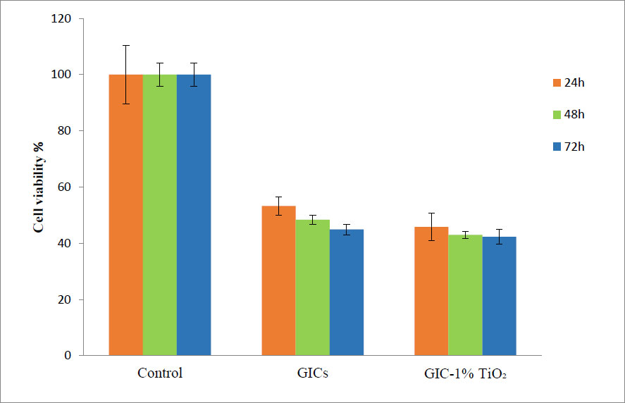
Bar graph illustrating the percentage viability of cells exposed to control, unmodified GIC, and GIC containing 1% TiO2 NPs after 24, 48, and 72 hours of treatment.
| Group | 24 h | 48 h | 72 h |
|---|---|---|---|
| Control (untreated) | 100.0 ± 2.0 | 100.0 ± 2.5 | 100.0 ± 2.2 |
| GIC (0 wt% TiO2) | 53.26 ± 3.3 | 48.35 ± 3.5 | 44.95 ± 2.9 |
| TiO2-GIC (1 wt% TiO2) | 45.82 ± 4.9 | 42.99 ± 1.34 | 42.33 ± 2.6 |
Note: Viability expressed as % of control.
| Source | df | F | p-value |
|---|---|---|---|
| Group (Control vs GIC vs TiO2-GIC) | 2 | 512.4 | <0.001 |
| Time (24 vs 48 vs 72 h) | 2 | 0.36 | 0.704 |
| Group × Time | 4 | 0.22 | 0.927 |
| Comparison | Mean Difference (%) | p-value | Interpretation |
|---|---|---|---|
| Control vs GIC | 42.5 | <0.001 | Significant |
| Control vs TiO2-GIC | 41.9 | <0.001 | Significant |
| GIC vs TiO2-GIC | 0.6 | 0.876 | Not significant |
The data demonstrate the impact of different materials and conditions on cellular inflammatory responses, as quantified by the levels of IL-6 and TNF-α cytokines (Table 4). In the negative control group, cytokine levels remained at baseline (IL-6: undetectable; TNF-α: 2.9 ± 2 pg/mL), indicating minimal inflammation. Exposure to LPS significantly elevated both IL-6 (163.1 ± 8 pg/mL) and TNF-α (14 ± 3 pg/mL) levels (p < 0.001 vs. negative control), confirming robust immune activation. Notably, when LPS was combined with conventional GIC, IL-6 levels increased further (194.9 ± 9 pg/mL), exceeding those observed with LPS alone (p < 0.01), suggesting that GIC may intensify the inflammatory response. In contrast, incorporation of 1% TiO2 into GIC significantly reduced IL-6 (36.2 ± 2 pg/mL) and TNF-α (9.2 ± 2 pg/mL) levels compared to GIC with LPS (p < 0.01), indicating a potential modulatory effect. Under non-stimulated conditions, both GIC and TiO2-GIC induced only minimal cytokine production (IL-6: 6.5 ± 3 and 4.6 ± 1 pg/mL; TNF-α: 2.2 ± 1 and 1.9 ± 1 pg/mL, respectively), with no significant differences from baseline (p > 0.05). These findings suggest that TiO2 incorporation may attenuate inflammation in activated environments without compromising baseline biocompatibility. Tables 5 to 7 represent the statistical results for cellular inflammatory responses.
| Conditions | IL-6 (pg/ml) | TNF-α (pg/ml) | Interpretation |
|---|---|---|---|
| Negative control | ND | 2.9±2 | Baseline inflammation |
| Positive control (+ LPS) | 163.1±8 | 14±3 | Strong inflammatory response due to LPS |
| TiO2-GIC + LPS | 36.2±2 | 9.2±2 | Reduced inflammation compared to GIC + LPS |
| GIC + LPS | 194.9±9 | 12.9±3 | Highest IL-6 response, strong inflammation |
| TiO2-GIC without LPS | 4.6±1 | 1.9±1 | Minimal inflammation, close to baseline |
| GIC without LPS | 6.5±3 | 2.2±1 | Slightly elevated IL-6, but still low |
Abbreviation: ND: Not Detected.
| Outcome | df (between, within) | F | p-value |
|---|---|---|---|
| IL-6 | 5, 12 | 1380.6 | <0.001 |
| TNF-α | 5, 12 | 18.7 | <0.001 |
| Comparison | Mean Difference (pg/mL) | 95% CI | p-value | Interpretation |
|---|---|---|---|---|
| LPS vs Negative control | +163.1 | 148.2 to 178.0 | <0.001 | Significant |
| GIC + LPS vs LPS | +31.8 | 18.6 to 45.0 | <0.01 | Significant |
| TiO2-GIC + LPS vs LPS | −126.9 | −141.4 to −112.3 | <0.001 | Significant |
| TiO2-GIC + LPS vs GIC + LPS | −158.7 | −173.1 to −144.2 | <0.001 | Significant |
| GIC without LPS vs Negative control | +6.5 | −2.1 to 15.1 | 0.281 | Not significant |
| TiO2-GIC without LPS vs Negative control | +4.6 | −3.8 to 13.0 | 0.392 | Not significant |
| GIC without LPS vs TiO2-GIC without LPS | +1.9 | −6.4 to 10.2 | 0.781 | Not significant |
| Comparison | Mean Difference (pg/mL) | 95% CI | p-value | Interpretation |
|---|---|---|---|---|
| LPS vs Negative control | +11.1 | 6.4 to 15.8 | <0.001 | Significant |
| GIC + LPS vs LPS | −1.1 | −4.2 to 2.0 | 0.612 | Not significant |
| TiO2-GIC + LPS vs LPS | −4.8 | −8.1 to −1.5 | <0.05 | Significant |
| TiO2-GIC + LPS vs GIC + LPS | −3.7 | −7.0 to −0.4 | <0.05 | Significant |
| GIC without LPS vs Negative control | −0.7 | −3.2 to 1.8 | 0.733 | Not significant |
| TiO2-GIC without LPS vs Negative control | −1.0 | −3.5 to 1.5 | 0.648 | Not significant |
| GIC without LPS vs TiO2-GIC without LPS | +0.3 | −2.1 to 2.7 | 0.942 | Not significant |
4. DISCUSSION
Dental restorative biomaterials are known to elicit both direct and indirect biological interactions with adjacent oral tissues. Numerous studies have investigated the cytotoxic potential of these materials, particularly their impact on fibroblast cell viability [4, 7, 27].
In the present study, HGFs were employed to evaluate the cytotoxicity of GIC. HGFs are considered highly suitable for such assessments due to ease of isolation from patients, rapid proliferation in culture, and high sensitivity in cytotoxicity assays [28]. The MTT assay was selected for cell viability evaluation. This colorimetric assay is widely recognized for its sensitivity, reliability, and quantitative accuracy in cytotoxicity testing of dental materials. Its rapid execution and cost-effectiveness further support its use in experimental studies [14]. TiO2 nanoparticles were incorporated at 1 wt% to achieve biological modulation while minimizing nanoparticle agglomeration and avoiding disruption of the acid–base setting reaction of glass ionomer cement. Higher loadings were avoided due to the potential for particle clustering, altered ion-release patterns, and undesirable changes in cement handling and matrix integrity [29].
Based on the findings of this study, incorporation of TiO2 NPs into GIC did not significantly alter cytotoxicity at varying time points. Modified GIC containing TiO2 NPs exhibited comparable cytotoxic profiles to conventional formulations. These results align with a previous report indicating that TiO2 NPs are non-cytotoxic [30]. In another investigation, the incorporation of TiO2 nanotubes into conventional GIC did not adversely affect its biological properties [31].
In the present study, both unmodified and TiO2 NP-modified GICs exhibited cytotoxic effects on HGFs within the first 24 hours of exposure. This observation aligns with earlier findings on HGFs [15] and mouse fibroblast L929 cells [16]. The observed cytotoxicity at 24 hours may be attributed to the release of metal ions and acidic by-products during the initial setting and maturation phases of the cement. The biocompatibility of dental materials is known to be influenced by their solubility and chemical composition [16]. In conventional GICs, cytotoxicity has been linked to the release of various metal ions, including iron [32], aluminum, zinc, and strontium [33]. Additionally, a strong correlation has been reported between fluoride release and cytotoxic responses in GICs [17, 34]. In an investigation, polyacrylic acid, the liquid constituent of GICs, was recognized as a principal factor contributing to cytotoxicity [35]. These acids, which are incorporated to enhance handling properties and reduce setting time, may possess irritant potential and negatively impact the overall biocompatibility of the material [36]. In contrast to the findings of the present study, a previous investigation reported that conventional GIC did not exhibit significant cytotoxicity toward HGFs within the first 24 hours, as assessed by the MTT assay [18]. Similarly, another study reported that conventional GIC was non-cytotoxic after 24 hours of exposure [17]. These discrepancies may be attributed to differences in the type of GICs used and the cellular models employed. While the current study utilized HGF cells, the aforementioned study used odontoblast-like MDPC-23 cells, which may respond differently to material exposure. The chemical composition and filler content of GICs are critical factors influencing cytotoxicity outcomes [7]. The GIC used in the present study was a restorative formulation, whereas the previous study [17] employed a luting GIC. Restorative GICs typically contain a higher filler load to enhance mechanical properties [7], which may contribute to increased cytotoxicity compared to luting variants.
Another finding of the present study was that cytotoxicity levels remained relatively unchanged over time. From 24 to 48 and 72 hours, all GIC formulations, regardless of TiO2 NPs incorporation, continued to exhibit cytotoxic effects. This suggests that the initial cytotoxic response persists over time and is not significantly mitigated by prolonged incubation. In this regard, previous studies have reported that conventional GICs exhibit cytotoxicity at 72 hours [19, 20]. It has also been shown that GICs may become non-cytotoxic after one week [34], likely due to cement maturation, pH normalization, and reduced ion release. Contrary to the results of the present study, one investigation observed a progressive decline in cell viability from 24 to 48 and 72 hours [15]. This discrepancy may be attributed to differences in the cellular models used; the above-mentioned study employed mouse fibroblast L929 cells, whereas the current study utilized HGFs. Furthermore, several studies reported a time-dependent enhancement in cell viability, observed between 24 and 72 hours of incubation [21, 22, 31]. The observed discrepancies in findings may be attributed to variations in cell type [21, 22], the nature of the tested materials [21], and the physicochemical form of the TiO2 NPs [31].
In the current study, two inflammatory mediators, including TNF-α and IL-6, were examined. TNF-α is one of the most critical proinflammatory cytokines that not only participates in vasodilation and blood coagulation regulation but also plays a role in increasing the synthesis of anti-inflammatory factors, such as IL-10 [23]. In a previous study, the anti-inflammatory effects of various restorative materials, including composite resin, compomer, and high-viscosity GIC, on L929 fibroblast cells were investigated. The findings demonstrated that these materials effectively reduced levels of TNF-α, IL-1, and IL-6, as measured by ELISA [23]. The results of our study demonstrated a modulation of the inflammatory response depending on the presence of LPS and the composition of the GIC. LPS alone significantly elevated both IL-6 and TNF-α levels, confirming its role as a potent proinflammatory stimulus. Interestingly, when LPS was combined with conventional GIC, IL-6 levels increased even further, suggesting that GIC may intensify the immune response under inflammatory conditions. This could be due to surface properties or leachable components that interact with immune cells. However, the inclusion of TiO2 NPs in GIC markedly reduced cytokine levels in the presence of LPS, indicating a potential anti-inflammatory or immunomodulatory effect. TiO2 NPs are known to influence cellular signaling, and in this context, they may be mitigating the proinflammatory cascade triggered by LPS. In the absence of LPS, both GIC formulations induced minimal cytokine release, reinforcing their biocompatibility under non-stimulated conditions.
Overall, these findings suggest that while conventional GIC may exacerbate inflammation when challenged with bacterial components, such as LPS, the incorporation of TiO2 NPs could improve their biological profile by attenuating this response. This has important implications for clinical applications, particularly in environments prone to bacterial exposure, such as dental restorations or orthopedic interfaces. Further investigation into the mechanisms by which TiO2 modulates immune signaling could support its use in designing next-generation biomaterials with enhanced immunological safety. Given that the present investigation was conducted under in vitro conditions, the interpretation of results should be approached with caution. It is well-recognized that in vitro outcomes may not fully reflect clinical performance [18]. Consequently, further research, particularly in vivo studies, is warranted to substantiate and expand upon these findings. In this study, only a single concentration of TiO2 NPs was assessed. Evaluating a broader range of concentrations would offer deeper insights into potential dose-dependent effects. The relatively short duration of the study also limits conclusions regarding long-term biological performance; thus, extended evaluations are recommended to determine the sustained efficacy of the modified GIC. The findings of the present study indicate that TiO2 NPs may enhance the biological profile of GIC without compromising cytocompatibility. However, further optimization is required to balance immunological safety with therapeutic efficacy. Future research should focus on refining nanoparticle formulations and exploring synergistic additives to improve both the mechanical and biological properties of restorative biomaterials.
CONCLUSION
This study examined the biological impact of incorporating TiO2 NPs into conventional GIC. The results demonstrated that TiO2-modified GIC exhibited comparable cell viability to the unmodified formulation. Importantly, the inclusion of TiO2 nanoparticles significantly reduced the expression of proinflammatory cytokines IL-6 and TNF-α, suggesting a potential immunomodulatory benefit.
AUTHORS’ CONTRIBUTIONS
The authors confirm their contributions to the paper as follows: Z.G.: Contributed to conceptualization, investigation, data curation, and writing of the original draft; Z.A.: Contributed to methodology, investigation, and writing of the original draft; S.K.: Contributed to supervision, data curation, validation, and review and editing of the manuscript; S.S.: Contributed to supervision, data curation, validation, writing of the original draft, and review and editing of the manuscript; S.M.D.: Contributed to investigation and writing of the original draft; E.J.N.: Contributed to the investigation; M.E.E.C.: Contributed to resources and investigation; F.D.T.: Contributed to the investigation.
LIST OF ABBREVIATIONS
| LPS | = Lipopolysaccharide |
| GICs | = Glass Ionomer Cements |
| FBS | = Fetal Bovine Serum |
| ELISA | = Enzyme-linked Immunosorbent Assay |
ETHICAL STATEMENT
This is an in vitro experimental study, for which ethical approval was granted by the Ethics Committee of Tabriz University of Medical Sciences, Iran (IR.TBZMED.VCR. REC.1403.065), approval date: 2024-05-20.
AVAILABILITY OF DATA AND MATERIALS
The data and supportive information is available within the article.
FUNDING
This research was financially supported by the Vice Chancellor for Research at Tabriz University of Medical Sciences, Iran and the authors gratefully acknowledge this support (Grant No. 73678).
CONFLICT OF INTEREST
Dr. Simin Sharifi is an Editorial Advisory Board member of The Open Dentistry Journal.
ACKNOWLEDGEMENTS
Declared none.